. 2010 Jun;19(6):1213-21.
doi: 10.1002/pro.400.

Dissecting the cAMP-inducible allosteric switch in protein kinase A RIalpha

Affiliations

Dissecting the cAMP-inducible allosteric switch in protein kinase A RIalpha

Timothy J Sjoberg et al. Protein Sci. 2010 Jun.

Abstract

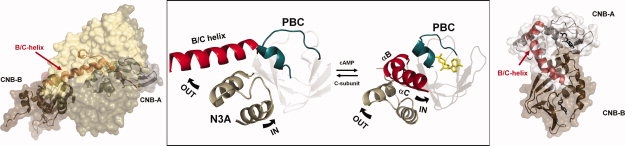
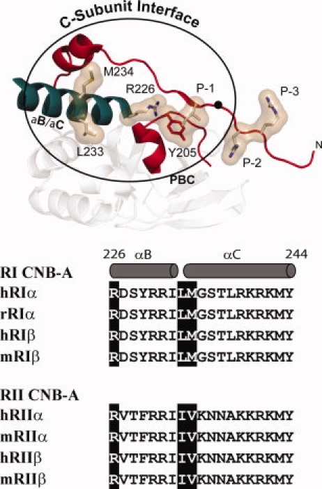
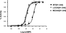
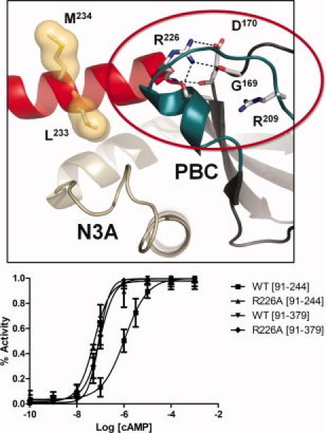
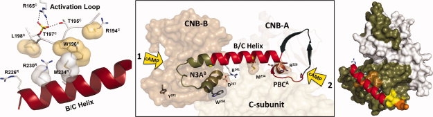
The regulatory subunits of cAMP-dependent protein kinase (PKA) are the major receptors of cAMP in most eukaryotic cells. As the cyclic nucleotide binding (CNB) domains release cAMP and bind to the catalytic subunit of PKA, they undergo a major conformational change. The change is mediated by the B/C helix in CNB-A, which extends into one long helix that now separates the two CNB domains and docks onto the surface of the catalytic subunit. We explore here the role of three key residues on the B/C helix that dock onto the catalytic subunit, Arg226, Leu233, and Met 234. By replacing each residue with Ala, we show that each contributes significantly to creating the R:C interface. By also deleting the second CNB domain (CNB-B), we show furthermore that CNB-B is a critical part of the cAMP-induced conformational switch that dislodges the B/C helix from the surface of the catalytic subunit. Without CNB-B the K(a) for activation by cAMP increases from 80 to 1000 nM. Replacing any of the key interface residues with Ala reduces the K(a) to 25-40 nM. Leu233 and M234 contribute to a hydrophobic latch that binds the B/C helix onto the large lobe of the C-subunit, while Arg226 is part of an electrostatic switch that couples the B/C helix to the phosphate binding cassette where the cAMP docks.


Figures

Figure 1
Figure 1
Conformational changes of the B/C helix in the CNB A domain of RIα. The major changes in conformation in RIα when the cAMP-bound conformation (right) is compared with the holoenzyme (left) are mediated by the unusual dynamics of the B/C helix show in red. In the center are highlighted the three conserved motifs in the CNB domain. In blue is the PBC, in tan is the N3A motif, and in red is the B/C helix. While the PBC, which is embedded in the middle of the stable β sandwich moves slightly, the helical subdomain undergoes major changes. Most significant is the extension of the B/C helix. Structures are from PDB: 1RGS and 2QCS.
Figure 2
Figure 2
C-Subunit interface in the CNB-A domain of RIα and sequence alignment of B/C helix. Top: Structure of the hydrophobic interface that is created by the conformational changes in CNB-A is shown. The hydrophobic residues that dock onto the C-subunit are highlighted. B/C helix is shown in teal while the PBC and the ordered linker and inhibitory site (residues 92–113 are shown in red). Bottom: Sequence comparison of the B/C helices in the four R-subunit isoforms. Residues mutated in this study are highlighted.
Figure 3
Figure 3
Effect of mutating Leu233 and Met234 in RIα(91–244) on activation by cAMP. Holoenzyme (25 nM) was incubated with increasing concentrations of cAMP and catalytic activity was measured. Catalytic activity was measured using a coupled Kemptide assay.
Figure 4
Figure 4
Activation of the Arg226Ala mutant by cAMP. The top panel shows the interactions of Arg226 in the holoenzyme structure of RIα(91–379):C:Mn2ATP (2QCS). The bottom panel shows activation of R226A holoenzyme by cAMP. Holoenzyme (25 nM) was formed with wild type and mutant forms of RIα(91–244) and RIα(91–379) and then incubated with increasing concentrations of cAMP (lower panel).
Figure 5
Figure 5
Both ends of the B/C helix are regulated by cAMP. The first cAMP binds to the B domain and helps to unleash the B/C helix from the large lobe of the C-subunit. This allows a second molecule of cAMP to bind to domain A, and this second cAMP is what disrupts the inhibitor peptide. This highly ordered and bivalent interaction of two molecules of cAMP with the B/C helix (center panel) provides a concerted allosteric mechanism for activation and release of kinase inhibition. The panel on the right shows how the B/C helix is anchored onto the large lobe of the C-subunit while the panel on the left shows how this B/C helix is buttressed up against Trp196 and Leu198 in the activation loop. Leu198 is the major determinant for the P + 1 docking site, and these two residues, Leu198 and Trp196, flank the essential phospho-Thr197 which is anchored to the active site through its interactions with Arg165 in the HRD motif in the catalytic loop.

Similar articles

Cited by

References

    1. Berman HM, Ten Eyck LF, Goodsell DS, Haste NM, Kornev A, Taylor SS. The cAMP binding domaan ancient signaling module. Proc Natl Acad Sci USA. 2005;102:45–50. - PMC - PubMed
    1. Canaves JM, Taylor SS. Classification and phylogenetic analysis of the cAMP-dependent protein kinase regulatory subunit family. J Mol Evol. 2002;54:17–29. - PubMed
    1. Kannan N, Wu J, Anand GS, Yooseph S, Neuwald AF, Venter JC, Taylor SS. Evolution of allostery in the cyclic nucleotide binding module. Genome Biol. 2007;8:R264. - PMC - PubMed
    1. Gill GN, Garren LD. Role of the receptor in the mechanism of action of adenosine 3′:5′-cyclic monophosphate. Proc Natl Acad Sci USA. 1971;68:786–790. - PMC - PubMed
    1. Ho RJ, Sutherland EW. Action of feedback regulator on adenylate cyclase. Proc Natl Acad Sci USA. 1975;72:1773–1777. - PMC - PubMed

Publication types

MeSH terms

Substances