. 2010 Jun;19(6):1253-7.
doi: 10.1002/pro.385.

Crystal structure of the crenarchaeal conserved chromatin protein Cren7 and double-stranded DNA complex

Affiliations

Crystal structure of the crenarchaeal conserved chromatin protein Cren7 and double-stranded DNA complex

Yingang Feng et al. Protein Sci. 2010 Jun.

Abstract

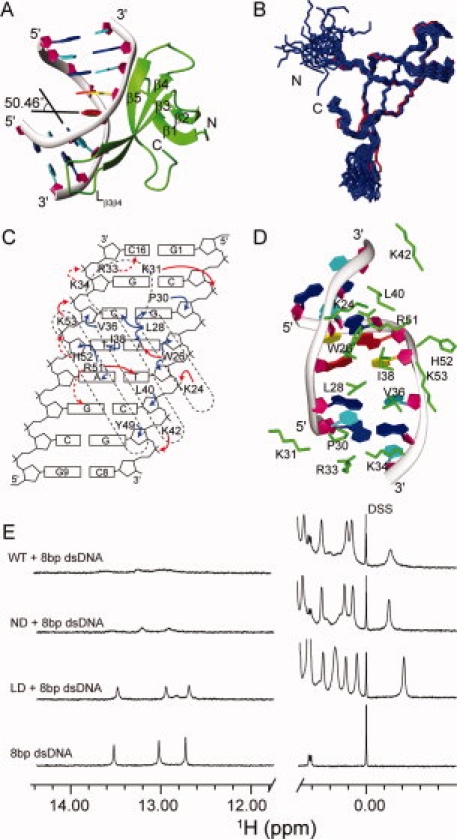
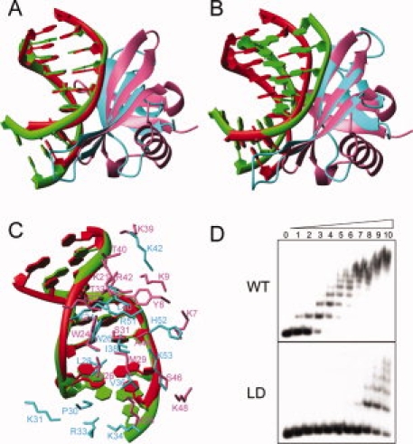
Cren7 is a crenarchaeal conserved chromatin protein discovered recently. To explore the mechanism of the DNA packaging in Crenarchaeota, the crystal structure of Cren7-GCGATCGC complex has been determined and refined at 1.6 A resolution. Cren7 kinks the dsDNA sharply similar to Sul7d, another chromatin protein existing only in Sulfolobales, which reveals that the "bending and unwinding" compacting mechanism is conserved in Crenarchaeota. Significant structural differences are revealed by comparing both protein-dsDNA complexes. The kinked sites on the same dsDNA in the complexes with Sul7d and Cren7 show one base pair shift. For Cren7, fewer charged residues in the beta-barrel structural region bind to DNA, and additionally, the flexible loop L(beta3beta4) is also involved in the binding. Electrophoretic mobility shift assays indicate that loop L(beta3beta4) is essential for DNA-binding of Cren7. These differences provide insight into the functional difference of both chromatin proteins, suggesting that Cren7 may be more regulative than Sul7d in the DNA-binding affinity by the methylation in the flexible loop L(beta3beta4) in vivo.


Figures

Figure 1
Figure 1
Crystal structure and protein–DNA interactions of the Cren7–dsDNA complex. A: Crystal structure of the Cren7–dsDNA complex. Cren7 is shown as green ribbon. The backbone of dsDNA is shown as gray ribbon, and the sugar ring is depicted as filled purple pentagon; A, T, C, G bases are colored in red, yellow, cyan, and blue, respectively. B: Superimposition of Cren7 in the complex (red) with the NMR solution structures of Cren7 (blue) (PDB: 2JTM). C: Schematic diagram summarizing all Cren7–DNA contacts. The filled red, filled blue, and dashed red arrows represent direct hydrogen bonds/salt bridges, van der Waals close contacts, and potential hydrogen bonds/salt bridges, respectively. D: Protein–DNA interactions in the structure. The side chains of residues involved in the interactions are shown as green sticks. E: 1D 1H NMR spectra of the Cren7–dsDNA complexes. The high field signals near 0.00 ppm are from methyl group of proteins, and the low field signals near 13.00 ppm are from dsDNA hydrogen-bonded base pairs. Full length Cren7 (WT) and Cren7ND show similar NMR spectra in the protein–dsDNA complexes. The peaks near 13.00 ppm are broadened extremely, indicating that almost all the dsDNA molecules form the complexes with Cren7 (WT). The spectrum of Cren7LD with dsDNA shows only slightly broadened peaks compared with those of the free dsDNA near 13.00 ppm, which indicates that the dsDNA in the Cren7LD-dsDNA complex is in a small fraction and fast exchange with the free dsDNA. An interactive view is available in the electronic version of the article. PRO385 Figure 1
Figure 2
Figure 2
Comparison of Cren7–dsDNA and Sul7d–dsDNA (PDB: 1AZP) complexes. A: Superimposition of dsDNA in the two complexes. Structures are superimposed using the backbone phosphor atoms of G3-C8 and C10-G15 in Cren7–dsDNA complex and C2-G7 and G11-C16 in Sul7d–dsDNA complex. The dsDNA and protein in the Cren7–dsDNA complex are colored in green and cyan, respectively. The dsDNA and protein in the Sul7d–dsDNA complex are colored in red and pink, respectively. B: Superimposition of proteins in the two complexes using the backbone atoms of the interface secondary structure residues (Cre7: 24–28, 36–42, 49–53; Sul7d, 22–26, 29–35, 40–44). C: Protein–DNA interactions in the both complexes superimposed as (A). D: The electrophoretic mobility shift assays of wild-type and loop-deletion mutant Cren7LD binding with a 60 bp dsDNA. Protein concentrations were 0, 0.02, 0.04, 0.08, 0.16, 0.31, 0.63, 1.3, 2.5, 5, and 10 μM, respectively. The apparent dissociation constants are ∼0.08 μM and 5 μM for wild-type Cren7 and Cren7LD, respectively. An interactive view is available in the electronic version of the article. PRO385 Figure 2

Similar articles

Cited by

References

    1. Luijsterburg MS, White MF, van Driel R, Dame RT. The major architects of chromatarchitectural proteins in bacteria, Archaea and eukaryotes. Crit Rev Biochem Mol Biol. 2008;43:393–418. - PubMed
    1. Luger K, Mader AW, Richmond RK, Sargent DF, Richmond TJ. Crystal structure of the nucleosome core particle at 2.8 A resolution. Nature. 1997;389:251–260. - PubMed
    1. Sandman K, Reeve JN. Archaeal chromatin proteins: different structures but common function? Curr Opin Microbiol. 2005;8:656–661. - PubMed
    1. Reeve JN. Archaeal chromatin and transcription. Mol Microbiol. 2003;48:587–598. - PubMed
    1. Dame RT. The role of nucleoid-associated proteins in the organization and compaction of bacterial chromatin. Mol Microbiol. 2005;56:858–870. - PubMed

Publication types

MeSH terms

LinkOut - more resources