. 2010 May 28;38(4):590-602.
doi: 10.1016/j.molcel.2010.02.040.

SWI/SNF has intrinsic nucleosome disassembly activity that is dependent on adjacent nucleosomes

Affiliations

SWI/SNF has intrinsic nucleosome disassembly activity that is dependent on adjacent nucleosomes

Mekonnen Lemma Dechassa et al. Mol Cell. .

Abstract

The ATP-dependent chromatin remodeling complex SWI/SNF regulates transcription and has been implicated in promoter nucleosome eviction. Efficient nucleosome disassembly by SWI/SNF alone in biochemical assays, however, has not been directly observed. Employing a model system of dinucleosomes rather than mononucleosomes, we demonstrate that remodeling leads to ordered and efficient disassembly of one of the two nucleosomes. An H2A/H2B dimer is first rapidly displaced, and then, in a slower reaction, an entire histone octamer is lost. Nucleosome disassembly by SWI/SNF did not require additional factors such as chaperones or acceptors of histones. Observations in single molecules as well as bulk measurement suggest that a key intermediate in this process is one in which a nucleosome is moved toward the adjacent nucleosome. SWI/SNF recruited by the transcriptional activator Gal4-VP16 preferentially mobilizes the proximal nucleosome and destabilizes the adjacent nucleosome.


Figures

Figure 1
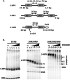
Figure 1. Remodeling of Short Oligonucleosomes with SWI/SNF
(A) Schematic representation of NPS used for di- and trinucleosome reconstitution is shown. NPS 601, 601b and 603 are the gray rectangles and the Gal4 binding site is an open rectangle. 2-NPS and 3-NPS refers to nucleosome reconstituted on two or three 601 NPS repeats; 2′-NPS, dinucleosome reconstituted on 601 (or 601b) and 603 NPS with Gal4 binding site. (B) Gel shift analysis is shown with mono-, di-and trinucleosomes remodeled with varying amounts of SWI/SNF and resolved by native 5% PAGE. NPS indicates the location of migration for the three different oligonucleosome substrates. The amounts of SWI/SNF were 3.5, 7, 14, 28, and 56 nM. The final concentration of nucleosomes based on octamer concentration was 45 nM and contained unlabeled plasmid DNA. The mononucleosome (1-NPS), dinucleosome (2-NPS) and trinucleosome (3-NPS) bands and corresponding DNA are indicated on the left side of the gel. In the labels on top of each gel the numbers refer to the length of DNA on either side of the positioned nucleosome, N, i.e., 81N6 is a mononucleosome flanked by 81 and 6 bp DNA. Vertical bars indicate remodeling products.
Figure 2
Figure 2. Effect of Linker Length on SWI/SNF Binding and Remodeling
(A) Dinucleosomes with 6, 30, and 79 bp of linker DNA were remodeled with different amounts of SWI/SNF and analyzed by native 5% PAGE under conditions similar to that in Figure 1. Remodeled dinucleosome products are indicated with a vertical bar corresponding to #1 or #2 and original nucleosomes as 2-NPS. (B) SWI/SNF was bound to dinucleosomes with different lengths of linker DNA and analyzed by native 4% PAGE. The SWI/SNF-bound dinucleosomes are indicated as SWI/SNF + 2 NPS and free dinucleosomes as 2-NPS.
Figure 3
Figure 3. Remodeling of Dinucleosomes by SWI/SNF Displaces One H2A/H2B Dimer and H3/H4 Tetramer
(A) Dinucleosomes (27N31N6; 2-NPS but without the Gal4 site) had DNA labeled with 32P and histone H2A or H3 of the octamer fluorescently labeled. The reactions contained 30 nM dinucleosomes with only 2-NPS DNA and 1.7, 5, or 15 nM SWI/SNF. After remodeling, the reactions were resolved by 4% native PAGE and visualized by scanning for fluorescent (upper panel) or radioactive (lower panel) signal. (B) Overlays of the H2A fluorescent (gray) and DNA radioisotope (32P-black) signals are shown with the signals normalized to the unremodeled dinucleosome. Left and right panels represent the overlays before and after SWI/SNF remodeling, respectively. (C) Same as (B) with fluorescent labeled H3 instead of H2A. (D-E) An analysis similar to that with dinucleosomes was also done with mononucleosomes (27N35) remodeled with SWI/SNF. Representative profiles of mononucleosome (27N35) assembled with octamers fluorescently labeled on C19 of H2A (D) and C80 of H3 (E) are shown. Left and right subpanels represent the overlays before and after SWI/SNF remodeling, respectively.
Figure 4
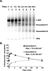
Figure 4. SWI/SNF Rapidly Displaces an H2A/H2B Dimer and in a Slower Step Displaces an Entire Nucleosome
(A) The rate at which the two species of remodeled dinucleosomes (same as in Figure 3) were formed was determined by gel shift assay. Remodeling reactions all contained ATP and were stopped by the addition of salmon sperm DNA and γ-S-ATP. The time points were 5, 15, and 45 sec; and 2, 6, 18 and 30 min. These reactions did not contain any DNA other than the 2-NPS DNA used to reconstitute the dinucleosomes the same as in Figure 3. (B) The relative amounts of remodeled species #1 (filled squares) and #2 (gray filled triangles) are plotted over time.
Figure 5
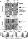
Figure 5. Site-Directed Mapping of Nucleosome Position Reveals that SWI/SNF Promotes Nucleosome Disassembly
(A) Nucleosome position was mapped by site-directed histone-DNA cross-linking and cleavage. Dinucleosomes were reconstituted on 2-NPS DNA (Figure 1A) using octamers containing cysteine at residue 120 of H3 and the reactions also contained salmon sperm carrier DNA. An overlay of the cleavage pattern of the dinucleosome is shown with the gray and black lines being respectively with and without SWI/SNF. The left portion of the overlay is horizontally expanded to better visualize cleavage products corresponding to the second nucleosome. Black bars highlight the new cleavage sites that occur after remodeling. The positions of nucleosomes after remodeling are schematically shown based on the new nucleosome positions observed. The nucleosomes are shown as ovals and DNA as white bars with the NPSs as black bars. The position of the pseudodyad axis is indicated with oval-shaped symbols and the cross-linking sites with vertical arrows. The direction of nucleosomes repositioning is shown with horizontal arrows. (B) Dinucleosomes reconstituted on 2′-NPS DNA (Figure 1A) using octamer with cysteine at residue 120 of H3 were used in site-directed histone-DNA contact mapping as in (A). Remodeling of dinucleosomes was conducted under conditions of limiting SWI/SNF with competitor DNA (sheared salmon sperm DNA) and was Gal4-VP16 dependent. Overlays of the cleavage pattern of the dinucleosome and corresponding schematic representation are displayed as in (A).
Figure 6
Figure 6. Mapping of Remodeled Single Dinucleosome Molecules Reveals that One of Two Nucleosomes Has Been Evicted
(A) Dinucleosomes were remodeled with SWI/SNF under non-recruited conditions as described in Figure 3 and did not contain carrier DNA. After remodeling SWI/SNF was competed off, and then the remodeled dinucleosomes were methylated by M.SssI, bisulfite treated, PCR amplified, and cloned into plasmid DNA. Individual clones were sequenced and regions protected against methylation are presented schematically by bars spanning at least two contiguously protected CG sites flanked by ≥ 2 methylated sites (i.e., one methylated site does not end a footprint). Methylated CG sites are shown with black dots, unmethylated CG with white dots. The data for free DNA, nucleosome only, and nucleosomes incubated with SWI/SNF in the absence of ATP are shown in Figure S3. The location of nucleosomes as determined by DNA methylation protection is schematically represented at right for dinucleosomes treated with SWI/SNF and ATP and placed into six different groups (1-6). The number of remodeled species in each group (in square brackets) and the percentage of all analyzed clones are shown at right. (B) Limiting amounts of SWI/SNF and competitor DNA were included in the reaction to make SWI/SNF binding and remodeling Gal4-VP16 dependent as in Figure 5B. Samples were processed for methylation footprinting as in (A). The data for free DNA, dinucleosomes only, dinucleosomes incubated with SWI/SNF and ATP and no Gal4-VP16, and dinucleosomes plus Gal4-VP16 are shown in Figure S4. The protection patterns were clustered into two groups (I and II). Chi-square analysis was used to test methylation differences between group I and II molecules at CpG sites in 601b NPS and 603 NPS as indicated by square brackets labeled with their respective highly significant P values. Schematics at right summarize nucleosome positions after remodeling with SWI/SNF recruited by Gal4-VP16 and are represented the same as in (A). Hatched circles indicate a nucleosome protection pattern that is more accessible than expected for canonical nucleosomes. (C) The percentage of DNA templates methylated at each of the 40 CpG sites is shown for the dinucleosome without (black bars) and with SWI/SNF and ATP added (gray bars). (D) The same type of analysis as in (C) is shown, except that it corresponds to SWI/SNF remodeling that is dependent on Gal4-VP16 as observed in (B).
Figure 7
Figure 7. Model for SWI/SNF-Mediated Disassembly of Nucleosomes in Arrays
(A) The approximate dimensions of SWI/SNF with respect to nucleosomes is depicted using two shades of blue for the two walls (front and back) that surround the trough of SWI/SNF where the nucleosome is thought to be bound. The location of the DNA translocase domain is shown as a filled black rectangle. The DNA initially associated with the nucleosome that is bound to SWI/SNF is highlighted in red; whereas the linker DNA and DNA associated with the other nucleosome are black and green, respectively. H2A/H2B (2A/2B) dimers and H3/H4 (3/4) tetramers in histone octamers (gray circle) are shown. The circled P and D refer to the proximal and distal nucleosomes, respectively. (B) Nucleosomal DNA is moved around the SWI/SNF-bound histone octamer by pulling linker DNA into the nucleosome. DNA bulges are created that propagate around the nucleosome and leave at the opposite nucleosome entry site. The second nucleosome is brought into close proximity to the SWI/SNF surface and can cause DNA to be displaced from the surface of one of the H2A/H2B dimers. (C) Loss of DNA contact with one H2A/H2B dimer promotes dissociation of the dimer from the histone octamer in a fast kinetic step. (D) Pulling more DNA away from the second nucleosome in order to move more DNA through the SWI/SNF-bound nucleosome is rate-limited by the stability of the interaction of the H3/H4 tetramer with DNA. Once DNA is transiently released from the tetramer SWI/SNF is poised to pull the extra DNA into and around the same SWI/SNF-bound nucleosome.

Comment in

References

    1. Adkins MW, Howar SR, Tyler JK. Chromatin disassembly mediated by the histone chaperone Asf1 is essential for transcriptional activation of the yeast PHO5 and PHO8 genes. Mol Cell. 2004;14:657–666. - PubMed
    1. Adkins MW, Williams SK, Linger J, Tyler JK. Chromatin disassembly from the PHO5 promoter is essential for the recruitment of the general transcription machinery and coactivators. Mol Cell Biol. 2007;27:6372–6382. - PMC - PubMed
    1. Almer A, Rudolph H, Hinnen A, Horz W. Removal of positioned nucleosomes from the yeast PHO5 promoter upon PHO5 induction releases additional upstream activating DNA elements. EMBO J. 1986;5:2689–2696. - PMC - PubMed
    1. Andrews AJ, Downing G, Brown K, Park YJ, Luger K. A thermodynamic model for Nap1-histone interactions. J Biol Chem. 2008;283:32412–32418. - PMC - PubMed
    1. Bazett-Jones DP, Côté J, Landel CC, Peterson CL, Workman JL. The SWI/SNF complex creates loop domains in DNA and polynucleosome arrays and can disrupt DNA-histone contacts within these domains. Mol Cell Biol. 1999;19:1470–1478. - PMC - PubMed

Publication types

MeSH terms

Substances

LinkOut - more resources