

Review
doi: 10.1016/j.cll.2010.01.004.
Hantaviruses
Affiliations
- PMID: 20513542
- PMCID: PMC2880890
- DOI: 10.1016/j.cll.2010.01.004
Item in Clipboard
Review
Hantaviruses
Clin Lab Med.
2010 Mar.
Abstract
Hantaviruses affect people worldwide, yet they remain poorly understood. This article explores the known history of hantaviruses. It describes diagnostic methods and potential options for treatment and prevention.
Published by Elsevier Inc.
Figures

Left panel shows the classification of negative stranded RNA viruses in two groups, Mononegavirales and segmented negative stranded RNA viruses. Mononegavirales have a single copy of negative sense RNA genome and segmented RNA viruses have multiple copies of negative sense RNA genome. Segmented RNA viruses have been further classified into three families, Orthomyxoviridae, Bunyaviridae and Arenaviridae. Viruses in the Bunyaviridae family have been classified into five genera, Bunyavirus, Hantavirus, Nairovirus, Phlebovirus and Tospovirus. Right panel show the rodent reservoirs for hantaviruses. (A) Apodemus agrarius (Reservoir for hantaan virus that cause HFRS); (B) deer mouse (Peromyscus maniculatus), (C) The Cotton Rat (Sigmodon hispidus); (D) The Rice Rat (Oryzomys palustris); (E) The White-footed Mouse (Peromyscus leucopus). Rodents in B, C, D and E cause HPS. All pictures in the right panel were obtained from the CDC web site; http://www.cdc.gov/NCIDOD/DISEASES/HANTA/HPS/noframes/rodents.htm
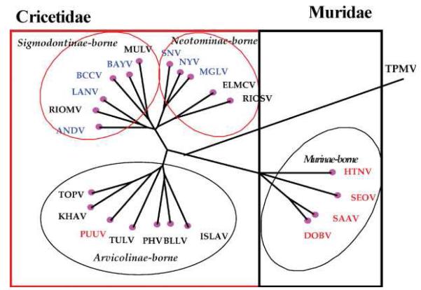
Phylogenic tree of hantaviruses carried by the different rodents (Family Muridae, subfamily Murinae and Family Cricetidae, subfamilies Arvicolinae, Sigmodontinae and Neotominae) and insectivores. The tree is based on the complete coding region of the S segment. HTNV, Hantaan virus; SEOV, Seoul virus; DOBV, Dobrava virus; SAAV, Saaremaa virus; PUUV, Puumala virus, TULV, Tula virus; PHV, Prospect Hill virus; BLLV, Blood Land Lake virus; ISLAV, Isla Vista virus; KHAV, Khabarovsk virus; TOPV, Topografov virus; SNV, Sin Nombre virus; NYV, New York virus; MGLV, Monongahela virus; ELMCV, El Moro Canyon virus; RIOSV, Rio Segundo virus; MULV, Muleshoe virus; BAYO, Bayou virus; BCCV, Black Creek Canal virus; LANV, Laguna Negra virus; RIOMV, Rio Mamore virus; ANDV, Andes virus; TPMV, Thottapalayam virus. In the figure, viruses causing HFRS are in red type and those causing HCPS in blue type. Viruses not associated with disease are in black type. This picture was obtained from reference (33)
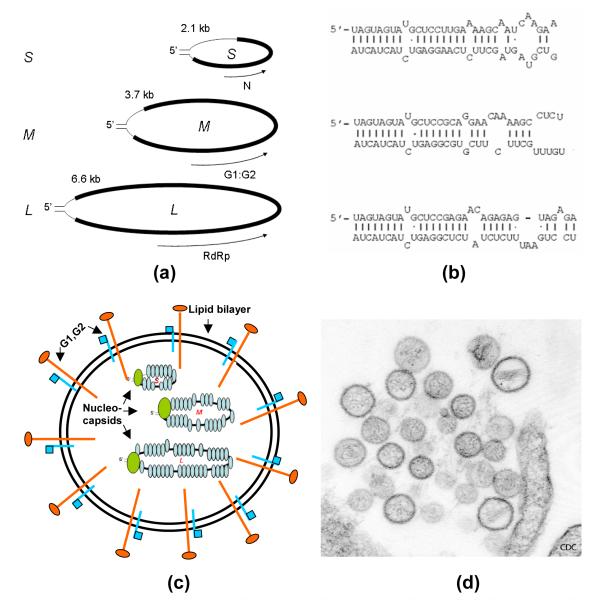
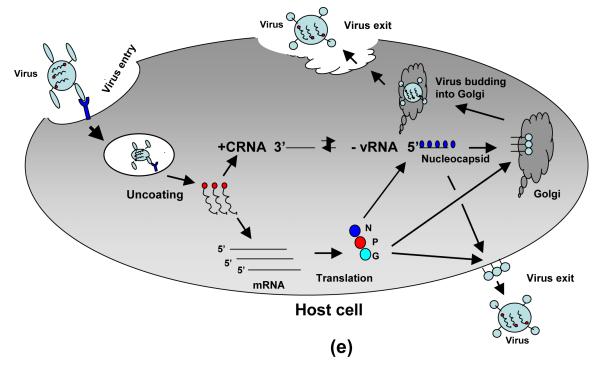
(a) Hantaviral genome comprises of three negative sense RNAs, S segment encodes nucleocapsid protein (N), M segment encodes glycoproteins G1 and G2, and L segment encodes viral RdRp. (b) Panhandle structures of three hantaviral genomic RNAs are formed by the base pairing of complementary bases at 5′ and 3′ terminus of each genome segment. (C) Pictorial representation of hantavirus particle, showing three nucleocapsids enveloped in a lipid bilayer. (d) Thin-section electron micrograph of Sin Nombre virus isolate, a causative agent of hantavirus pulmonary syndrome (HPS) http://www.cdc.gov/ncidod/diseases/hanta/hps/noframes/hpsem.htm . (e) Pictorial representation of hantavirus life cycle.
(a) Hantaviral genome comprises of three negative sense RNAs, S segment encodes nucleocapsid protein (N), M segment encodes glycoproteins G1 and G2, and L segment encodes viral RdRp. (b) Panhandle structures of three hantaviral genomic RNAs are formed by the base pairing of complementary bases at 5′ and 3′ terminus of each genome segment. (C) Pictorial representation of hantavirus particle, showing three nucleocapsids enveloped in a lipid bilayer. (d) Thin-section electron micrograph of Sin Nombre virus isolate, a causative agent of hantavirus pulmonary syndrome (HPS) http://www.cdc.gov/ncidod/diseases/hanta/hps/noframes/hpsem.htm . (e) Pictorial representation of hantavirus life cycle.
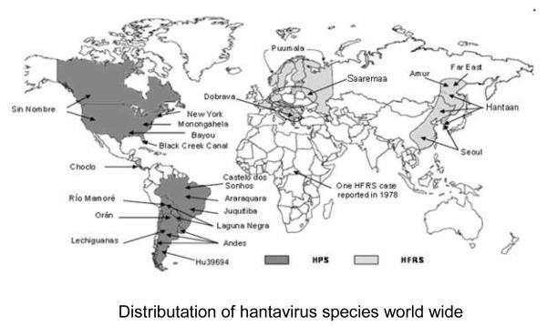
World wide distributation of hantavirus species
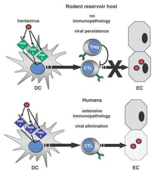
Working hypothesis of differential regulation of hantavirus-specific immune responses in rodent reservoir hosts and humans. During their encounter with viruses DCs integrate different signals received through several PRRs (PRR 1, PRR 2, PRR3, etc.) which determine the quality of the ensuing T-cell response. (A) In their rodent reservoir host, hantavirus-associated PRR signaling could program DCs to stimulate Treg cells that can suppress virus-specific CTLs, leading to viral persistence and at the same time preventing virus-induced immunopathology. (B) In humans, who are not adapted to hantaviruses, PRR signaling in DCs results in a dominant antiviral CTL response. As a consequence, hantavirus-infected endothelial cells (EC) are immediately eliminated leading to immunopathology. Note: This picture was obtained from Ref (29)
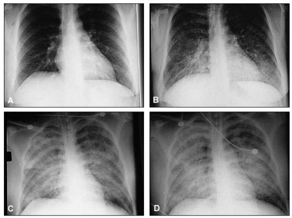
Radiographs showing the evolution of hantavirus pulmonary syndrome in a 30-year-old woman. (A) Chest radiograph before onset of illness. (B) Admission radiograph. (C) Radiograph after intubation. (D) Radiograph just before death. Note: This picture was obtained from Ref (11)
References
-
- Center for Disease Control and Prevention Case definitions for infectious conditions under public health surveillance. MMWR Morb Mortal Wkly Rep. 1997;46:1–55. - PubMed
-
- Banchereau J, Steinman RM. Dendritic cells and the control of immunity. Nature. 1998;392:245–52. - PubMed
-
- Bradley JR. TNF-mediated inflammatory disease. J Pathol. 2008;214:149–60. - PubMed
-
- Carey DE, Reuben R, Panicker KN, Shope RE, Myers RM. Thottapalayam virus: a presumptive arbovirus isolated from a shrew in India. Indian J Med Res. 1971;59:1758–60. - PubMed
-
- Duchin JS, Koster FT, Peters CJ, Simpson GL, Tempest B, Zaki SR, Ksiazek TG, Rollin PE, Nichol S, Umland ET, et al. Hantavirus pulmonary syndrome: a clinical description of 17 patients with a newly recognized disease. The Hantavirus Study Group. N Engl J Med. 1994;330:949–55. - PubMed
Publication types
MeSH terms
Grants and funding
LinkOut - more resources
Full Text Sources
Medical
