Review
. 2010 Mar;30(1):261-92.
doi: 10.1016/j.cll.2009.10.004.

Human ehrlichiosis and anaplasmosis

Affiliations
Review

Human ehrlichiosis and anaplasmosis

Nahed Ismail et al. Clin Lab Med. 2010 Mar.

Abstract

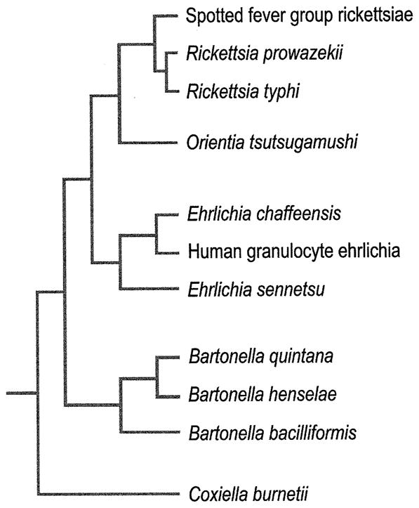
Human ehrlichiosis and anaplasmosis are acute febrile tick-borne diseases caused by various members of the genera Ehrlichia and Anaplasma (Anaplasmataceae). Human monocytotropic ehrlichiosis has become one of the most prevalent life-threatening tick-borne disease in the United States. Ehrlichiosis and anaplasmosis are becoming more frequently diagnosed as the cause of human infections, as animal reservoirs and tick vectors have increased in number and humans have inhabited areas where reservoir and tick populations are high. Ehrlichia chaffeensis, the etiologic agent of human monocytotropic ehrlichiosis (HME), is an emerging zoonosis that causes clinical manifestations ranging from a mild febrile illness to a fulminant disease characterized by multiorgan system failure. Anaplasma phagocytophilum causes human granulocytotropic anaplasmosis (HGA), previously known as human granulocytotropic ehrlichiosis. This article reviews recent advances in the understanding of ehrlichial diseases related to microbiology, epidemiology, diagnosis, pathogenesis, immunity, and treatment of the 2 prevalent tick-borne diseases found in the United States, HME and HGA.


Figures

Figure 1
Figure 1
Phylogenetic relationships between rickettsias based on 16S rRNA gene sequences. (From Mason PR, Kelly PJ. Ch. 235: Rickettsia and Rickettsia-Like Organisms. In: Cohen & Powderly, editors. Infectious Diseases, 2nd ed. Mosby; 2004. Permission requested from Elsevier.)
Figure 2
Figure 2
Peripheral blood leukocytes containing ehrlichial morula in patients with human monocytic ehrlichiosis (A) and human granulocytic anaplasmosis (B and C). A and B, a morula (arrow) containing Ehrlichia chaffeensis in a monocyte in patient with HME. B and C; a morula (arrowhead) containing Anaplasma phagocytophilum in a neutrophil in patient with HGA. Wright stains, original magnifications ×1,200. (A From Walker DH, Dumler JS. Ch. 190: Ehrlichia chaffeensis (Human Monocytotropic Ehrlichiosis), Anaplasma phagocytophilum (Human Granulocytotropic Anaplasmosis), and Other Ehrlichiae. In: Mandell, Bennett, & Dolin, editors. Principles and Practice of Infectious Diseases, 6th ed. Church Livingstone; 2005. Permission requested from Elsevier. B From Siberry GK, Dumler JS. Ch. 228: Ehrlichiosis and Anaplasmosis. In: Kliegman, editor. Nelson Textbook of Pediatrics, 18th ed. Saunders; 2007. Permission requested from Elsevier. C From Walker DH, Paddock CD, Dumler JS. Emerging and Re-emerging Tick-Transmitted Rickettsial and Ehrlichial Infections. Medical Clinics of North America, 2008; 92(6). Permission requested from Elsevier.)
Figure 3
Figure 3
Light microscopic picture of canine monocytes (DH82) are heavily infected in vitro with E. chaffeenesis (A) and E. canis (B). Typical ehrlichial inclusions (morulae) are present inside the cytoplasm of infected cells (Giemsa staining. Orginal magnification × 200. (C) Giemsa-stained ISE6 cells infected with A. phagocytophilum strain isolated from a female I. scapularis tick. Anaplasma organisms exist in large intracellular vacuoles (arrow). N, host cell nucleus. Giemsa stain. (A Courtesy of Ismail N. B From Mason PR, Kelly PJ. Ch. 235: Rickettsia and Rickettsia-Like Organisms. Cohen & Powderly: Infectious Diseases, 2nd ed. 2004; permission requested from Elsevier. C From Massung RF, Levin ML, Munderloh UG et al. Isolation and Propagation of the Ap-Variant 1 Strain of Anaplasma phagocytophilum in a Tick Cell Line. J of Clin Micro, 2007; 2138–2143. Permission requested from JCM.)
Figure 4
Figure 4. Electron micrographs of E. chaffeensis interaction with DH82 cells
(A) At 24h post-infection, a single reticulate cell (RC) divide by binary fission (black arrowhead). (B) At 48h post-infection, two morulae (black arrowheads) contain RC. (C) At 72 h post infection, Ehrlichia have matured into dense core (DC). Three morulae contain DC (black arrows), and one morula contains RC (black arrowhead). (D) High power of a morula containing DC at 72 h post infection. Mitochondria surrounding morulae were indicated by white arrows. (From Zhang J-Z, Popov VL, Gao S, et al. The developmental cycle of Ehrlichia chaffeensis in vertebrate cells. Cellular Microbiology, 2006; 9(3):610–618. Permission requested from Blackwell Publishing Ltd.)
Figure 5
Figure 5
Ehrlichia TR47: ultrastructure and confocal microscopy. Differential expression of TRP47 (red) on dense-cored E. chaffeensis cultured in vitro (DH82 cells) as visualized using three color scanning laser confocal fluorescent microscopy. E. chaffeensis infected cells were dually stained with rabbit anti-Ehrlichia disulfide bond formation protein (Dsb) (green-Alexa Fluor 488), and mouse anti-E. chaffeensis TRP47 (red-Alexa Fluor 568). Host cell nuclei were counterstained with 4′, 6′-diamidino-2-phenylindole, dihydrochloride (DAPI) and images merged. (Courtesy of McBride JW.)
Figure 6
Figure 6
A) Number of ehrlichiosis cases caused by E. chaffeensis reported to CDC by State Health Department from 1999–2006. B) Average reported annual incidence of human monocytic ehrlichiosis by state –United States, 2001–2002 and nationwide. (Open access: published by the Centers for Disease Control and Prevention, a U.S. Government agency and are in the public domain and can be used without permission.)
Figure 7
Figure 7
A) Lone Star tick; Amblyomma americanum that transmit the agent of human monocytic ehrlichiosis B) Life Cycle of monocytotropic E. chaffeensis. (Courtesy of Bloch KC, and open access: published by the Centers for Disease Control and Prevention, a U.S. Government agency and are in the public domain and can be used without permission)
Figure 8
Figure 8
A) Number of ehrlichiosis cases caused by A. phagocytophilum reported to CDC by State Health Department from 1999–2006. B) Average reported annual incidence of human monocytic ehrlichiosis by state –United States, 2001–2002 and nationwide. (Open access: published by the Centers for Disease Control and Prevention, a U.S. Government agency and are in the public domain and can be used without permission.)
Figure 9
Figure 9
A) Blacklegged tick; Ixodes Scapularis that transmit the agent of human granulocytic ehrlichiosis and Lyme disease. B) Western black-legged tick; Ixodes Scapularis that transmit the agent of human granulocytic ehrlichiosis (A & B Open access: published by the Centers for Disease Control and Prevention, a U.S. Government agency and are in the public domain and can be used without permission)
Figure 10
Figure 10
A) Worldwide distribution of Ehrlichia and Anaplasma. HGA: human granulocytic anaplasmosis; HME: human monocytic ehrlichiosis. B) Global health map shows worldwide distribution of HGA and HME marked by yellow dots. (A From Raoult D. Ch. 348: Rickettsioses. In Goldman, editor. Cecil Medicine, 23rd ed. Saunders, 2007. Permission requested from Elsevier. B From “Worldwide Outbreak of Ehrilichia” tracking map from http://www.healthmap.com [accessed June 15, 2009]. Permission granted.)
Figure 11
Figure 11
A) Hepatic histopathology in the murine model of fatal monocytic ehrlichiosis caused by systemic infection with virulent moncytic Ehrlichia (IOE). H&E staining shows extensive focal necrosis and apoptosis of hepatocytes (arrows). B) Hepatic histopathology in the murine model of mild monocytic ehrlichiosis caused by infection with mildly virulent Ehrlichia muris. H&E staining shows formation of well formed granuloma (arrowhead). (From Ismail N, Soong L, McBride JW, et al. Overproduction of TNF-alpha by CD8+ type 1 cells and down-regulation of IFN-gamma production by CD4+ Th1 cells contribute to toxic shock-like syndrome in an animal model of fatal monocytotropic ehrlichiosis. J Immunol, 2004; 172(3):1786–800. Permission granted.)
Figure 12
Figure 12
A) Hemophagocytosis (arrows) in lymph nodes from a patient with HME (H&E; original magnification 240×). B) High cellularity in lymph node from patient with HME (magnificationX 64). C) Immunohistochemical staining of lymph nodes from patient with HME shows typical low bacterial burden (immunoperoxidase with hematoxylin counterstain; original magnification 240×). (From Dierberg KL, Dumler JS. Lymph node hemophagocytosis in rickettsial diseases: a pathogenetic role for CD8 T lymphocytes in human monocytic ehrlichiosis (HME)? In: BMC Infect Dis. 2006; 6:121. This is an Open Access article distributed under the terms of the Creative Commons Attribution License (http://creativecommons.org/licenses/by/2.0), which permits unrestricted use, distribution, and reproduction in any medium, provided the original work is properly cited.)

References

    1. Dumler JS, Barbet AF, Bekker CP, et al. Reorganization of genera in the families Rickettsiaceae and Anaplasmataceae in the order Rickettsiales: Unification of some species of Ehrlichia with Anaplasma, Cowdria with Ehrlichia and Ehrlichia with Neorickettsia, descriptions of six new species combinations and designation of Ehrlichia equi and ‘HGE agent’ as subjective synonyms of Ehrlichia phagocytophila. Int J Syst Evol Microbiol. 2001;51:2145–2165. - PubMed
    1. Dawson JE, Anderson BE, Fishbein DB, et al. Isolation and characterization of an Ehrlichia sp. from a patient diagnosed with human ehrlichiosis. J Clin Microbiol. 1991;29:2741–2745. - PMC - PubMed
    1. Dumler JS, Bakken JS. Ehrlichial diseases of humans: emerging tick-borne infections. Clin Infect Dis. 1995;20:1102–1110. - PubMed
    1. Anderson BE, Dawson JE, Jones DC, et al. Ehrlichia chaffeensis, a new species associated with human ehrlichiosis. J Clin Microbiol. 1991;29:2838–2842. - PMC - PubMed
    1. Paddock CD, Liddell AM, Storch GA. Other causes of tick-borne ehrlichioses, including Ehrlichia ewingii. In: Goodman JL, Dennis DT, Sonenshine DE, editors. Tick-borne diseases of humans. ASM Press; Washington DC: 2005. pp. 258–267.

Publication types