. 2010 May 31;189(5):777-82.
doi: 10.1083/jcb.201004104.

Metadata matters: access to image data in the real world

Affiliations

Metadata matters: access to image data in the real world

Melissa Linkert et al. J Cell Biol. .

Abstract

Data sharing is important in the biological sciences to prevent duplication of effort, to promote scientific integrity, and to facilitate and disseminate scientific discovery. Sharing requires centralized repositories, and submission to and utility of these resources require common data formats. This is particularly challenging for multidimensional microscopy image data, which are acquired from a variety of platforms with a myriad of proprietary file formats (PFFs). In this paper, we describe an open standard format that we have developed for microscopy image data. We call on the community to use open image data standards and to insist that all imaging platforms support these file formats. This will build the foundation for an open image data repository.


Figures

Figure 1.
Figure 1.
Example data in the JCB DataViewer. An example of original image data associated with this paper, viewed in the JCB DataViewer. The image shows the following: a 3D stack of a fixed HeLa cell stained with DAPI (blue), anti-INCENP (red), and anti-tubulin (green), recorded using a wide-field microscope; a time-lapse video of a C. elegans embryo expressing GFP-tubulin, recorded using a multiphoton microscope; a transmission electron microscope (TEM) image of bacteriophages visualized using negative stain; a 3D stack of a fixed HeLa cell stained with anti-tubulin, recorded using an OMX 3D structured illumination microscope; a TEM image of Rb bound to DNA; and a 5D image of GFP-coilin and YFP-histone H2B in a HeLa cell, recorded by wide-field microscopy (Platani et al., 2000). An example view of metadata is included at the bottom left. Note that available metadata differ substantially between the different images, depending on the metadata that are stored in the original files. These images and their associated metadata are available at http://jcb-dataviewer.rupress.org/jcb/browse/2859/.
Figure 2.
Figure 2.
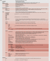
Recommendations for OME Compliant image metadata. The Image and Instrument Elements from the OME Data Model, with attributes and hierarchies shown in diagrammatic form. The Image Element contains core metadata that can be used for display and processing of the associated binary image data. Currently, an OME Compliant image completes all of the metadata in the Image Element. By the end of 2010, we aim to include the Instrument Element in the OME Compliant specification. The Bio-Formats library provides support for writing OME-XML either as a stand-alone file or within the header of an OME-TIFF file. The full XML Schema version of the OME Data Model is available at http://ome-xml.org/browser/Schemas/OME/2010-04/ome.xsd. Updates to the OME Data Model are announced on the project’s roadmap site (http://ome-xml.org/roadmap).

Similar articles

Cited by

References

    1. Christiansen J.H., Yang Y., Venkataraman S., Richardson L., Stevenson P., Burton N., Baldock R.A., Davidson D.R. 2006. EMAGE: a spatial database of gene expression patterns during mouse embryo development. Nucleic Acids Res. 34:D637–D641 10.1093/nar/gkj006 - DOI - PMC - PubMed
    1. Committee on Science, Engineering, and Public Policy (US), and Committee on Ensuring the Utility and Integrity of Research Data in a Digital Age 2009. Ensuring the Integrity, Accessibility, and Stewardship of Research Data in the Digital Age. National Academies Press, Washington, DC: 162 pp - PubMed
    1. Field D., Sansone S.A., Collis A., Booth T., Dukes P., Gregurick S.K., Kennedy K., Kolar P., Kolker E., Maxon M., et al. 2009. Megascience. ’Omics data sharing. Science. 326:234–236 10.1126/science.1180598 - DOI - PMC - PubMed
    1. Goldberg I.G., Allan C., Burel J.-M., Creager D., Falconi A., Hochheiser H.S., Johnston J., Mellen J., Sorger P.K., Swedlow J.R. 2005. The Open Microscopy Environment (OME) data model and XML file: open tools for informatics and quantitative analysis in biological imaging. Genome Biol. 6:R47 10.1186/gb-2005-6-5-r47 - DOI - PMC - PubMed
    1. Haudry Y., Berube H., Letunic I., Weeber P.D., Gagneur J., Girardot C., Kapushesky M., Arendt D., Bork P., Brazma A., et al. 2008. 4DXpress: a database for cross-species expression pattern comparisons. Nucleic Acids Res. 36:D847–D853 10.1093/nar/gkm797 - DOI - PMC - PubMed

Publication types

MeSH terms