. 2010 May 31;189(5):871-83.
doi: 10.1083/jcb.201001008.

Multiple mechanisms collectively regulate clathrin-mediated endocytosis of the epidermal growth factor receptor

Affiliations

Multiple mechanisms collectively regulate clathrin-mediated endocytosis of the epidermal growth factor receptor

Lai Kuan Goh et al. J Cell Biol. .

Abstract

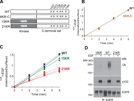
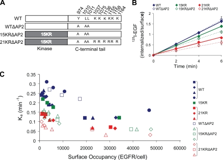
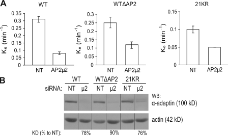
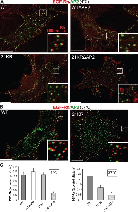
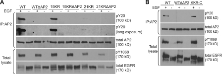
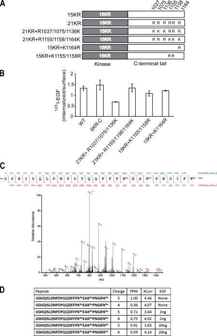
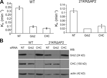
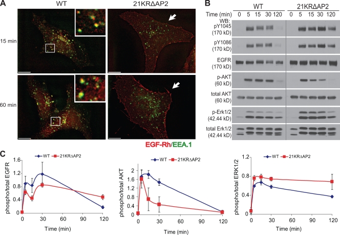
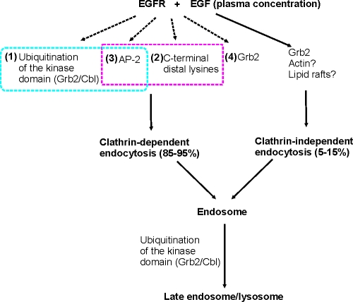
Endocytosis of the epidermal growth factor receptor (EGFR) is important for the regulation of EGFR signaling. However, EGFR endocytosis mechanisms are poorly understood, which precludes development of approaches to specifically inhibit EGFR endocytosis and analyze its impact on signaling. Using a combination of receptor mutagenesis and RNA interference, we demonstrate that clathrin-dependent internalization of activated EGFR is regulated by four mechanisms, which function in a redundant and cooperative fashion. These mechanisms involve ubiquitination of the receptor kinase domain, the clathrin adaptor complex AP-2, the Grb2 adaptor protein, and three C-terminal lysine residues (K1155, K1158, and K1164), which are acetylated, a novel posttranslational modification for the EGFR. Based on these findings, the first internalization-defective EGFR mutant with functional kinase and normal tyrosine phosphorylation was generated. Analysis of the signaling kinetics of this mutant revealed that EGFR internalization is required for the sustained activation of protein kinase B/AKT but not for the activation of mitogen-activated protein kinase.


Figures

Figure 1.
Figure 1.
Mutation of Lys residues in the C terminus decreases internalization of 125I-EGF by the 15KR mutant. (A) Schematic representation of 6KR-C and 21KR EGFR mutants in which six Lys were replaced by arginines in the C-terminal tail of wtEGFR and 15KR, respectively. (B) Time course of 1 ng/ml 125I-EGF internalization in PAE cells expressing wtEGFR (blue) or 6KR-C (orange) receptor. (C) Time course of 1 ng/ml 125I-EGF internalization in various single-cell clones of PAE cells expressing wtEGFR (blue), 15KR (green), or 21KR (red). (D) PAE cells expressing wtEGFR, 15KR, and 21KR were untreated or treated with 20 ng/ml EGF for 5 min at 37°C, and EGFR was immunoprecipitated (IP). Immunoprecipitates were probed by Western blotting with antibodies to ubiquitin (Ub), phosphotyrosine (pY20), and EGFR (1005). MW, molecular weight; WB, Western blot.
Figure 2.
Figure 2.
Mutation of AP-2–binding motifs (Y974RAL and LL1010/1011) decreases internalization of 125I-EGF by the 15KR and 21KR mutants. (A) Schematic representation of WTΔAP2, 15KRΔAP2, and 21KRΔAP2 EGFR mutants in which Tyr974 and LL1010/1011 were mutated to alanines in wtEGFR, 15KR, and 21KR mutants, respectively. (B) Summary of 1 ng/ml 125I-EGF internalization experiments performed in several single-cell clones of PAE cells expressing wtEGFR, WTΔAP2, 15KR, 15KRΔAP2, 21KR, or 21KRΔAP2 receptors. Bars represent SEM from three to four independent experiments. (C) Internalization saturation plots are shown. Specific internalization rate constant (ke) was measured in several clones of PAE cells expressing wtEGFR or various EGFR mutants treated with 1–20 ng/ml 125I-EGF. ke values are plotted against the number of surface EGFR occupied by 125I-EGF per cell measured as the amount of surface 125I-EGF after 5 min of continuous 125I-EGF internalization at 37°C.
Figure 3.
Figure 3.
siRNA depletion of AP-2 decreases internalization of 125I-EGF to a various degree in cells expressing wtEGFR, WTΔAP2, and 21KR receptors. (A) Internalization rate constants (ke) were measured using 1 ng/ml 125I-EGF in wtEGFR, WTΔAP2, and 21KR-expressing cells that were transfected with nontargeting siRNA (NT) or siRNA targeting µ2 subunit of AP-2. Bars represent SEM from four experiments. (B) Lysates of cells used in internalization experiments described in A were probed by Western blotting (WB) with Ab32 to β-adaptin and actin antibody (loading control). All images are from the same Western blot. The efficiency of siRNA depletion (KD%) was calculated as a percentage of the intensity of the β-adaptin signal in depleted cells to that intensity in nontargeting siRNA–transfected cells (both signals were first normalized to the loading control). Black lines indicate that intervening lanes have been spliced out.
Figure 4.
Figure 4.
Recruitment of EGF-Rh into clathrin-coated pits is impaired in cells expressing 21KR and 21KRΔAP2 mutants. (A) PAE cells expressing wtEGFR, WTΔAP2, 21KR, and 21KRΔAP2 were transiently transfected with pcDNA3.1–β2-YFP. After 2 d, the cells were incubated with 4 ng/ml EGF-Rh for 45 min at 4°C and fixed. A z stack of optical sections was acquired through CY3 (EGF-Rh) and FITC (β2-YFP) filter channels and deconvoluted. 21KRΔAP2 was mostly localized to the ruffles at cell periphery. (B) PAE cells expressing wtEGFR and 21KR were transiently transfected with pcDNA3.1–β2-YFP. After 2 d, the cells were incubated with 4 ng/ml EGF-Rh for 1 min at 37°C and fixed. A z stack of optical sections was acquired through CY3 (EGF-Rh) and FITC (β2-YFP) filter channels and deconvoluted. (A and B) Insets show enlarged views of boxed areas with CY3 filter channel shifted ∼300 nm to the right to better demonstrate the localization of EGF-Rh in coated pits. Bars, 10 µm. (C) At least 10 images obtained for each cell clone as described in A and B were used to quantitate the percentage of rhodamine fluorescence colocalized with the punctate β2-YFP fluorescence dots (coated pits). Error bars indicate SEM.
Figure 5.
Figure 5.
EGF-induced tyrosine phosphorylation of β2-adaptin is impaired in cells expressing 21KR and various AP-2–binding motif EGFR mutants. PAE cells expressing wtEGFR and various mutants were untreated or treated with 100 ng/ml EGF for 5 min, lysed, and AP-2 was immunoprecipitated (IP) with AP.6 antibody. Immunoprecipitates were blotted with phosphotyrosine antibody (pY20) and antibody to α-adaptin (AC.1). Overexposed blot demonstrates the presence of the weak pY20 signal in AP-2 immunoprecipitates from 21KR-expressing cells. Aliquots of lysates (bottom) were blotted with antibodies to phospho-Tyr1068 of EGFR and total EGFR (1005). Two representative experiments (A and B) are shown.
Figure 6.
Figure 6.
Three distal Lys in the C terminus of EGFR (K1155, K1158, and K1164) are important for internalization and are acetylated. (A) Schematic representation of 21KR + R1037/1075/1136K, 21KR + R1155/1158/1164K, 15KR + K1164R, and 15KR + K1155/1158R EGFR mutants. (B) Internalization rates of 1 ng/ml 125I-EGF are compared in cells expressing wtEGFR, 6KR-C, and mutants depicted in A. Error bars indicate mean values of ke (SEM) obtained in two individual single-cell clones for each mutant. (C) MS analysis of wtEGFR immunoprecipitates from PAE cells treated and untreated with EGF. Representative MS/MS spectrum of EGFR with acetylation (Ac) at Lys 1155, 1158, and 1164 and the sequence of the peptide. The precursor peptide ion (m/z 837.4064) was isolated and fragmented in a mass spectrometer. Fragment ions included both the N and C termini (b and y ions, respectively) in which the detected ions are underlined. (D) The ion charge state, mass deviation, Sequest score (XCorr), and experimental condition are shown for the peptides containing Lys1155, 1158, and 1164 that were identified. In addition, a tryptic peptide containing unmodified Lys1155 was identified multiple times (not depicted).
Figure 7.
Figure 7.
siRNA depletion of Grb2 and CHC decreases internalization of 125I-EGF in cells expressing wtEGFR and 21KRΔAP2 mutant. (A) Internalization rate constants (ke) were measured using 1 ng/ml 125I-EGF in wtEGFR and 21KRΔAP2-expressing cells that were transfected with nontargeting siRNA (NT) or siRNA targeting Grb2 or CHC. Error bars indicate SEM from three experiments. (B) Lysates of cells used in internalization experiments described in A were probed by Western blotting with antibodies to CHC, Grb2, and actin (loading control). All images are from the same Western blot (WB). Black lines indicate that intervening lanes have been spliced out.
Figure 8.
Figure 8.
Poor accumulation of 21KRΔAP2 mutant in endosomes and altered kinetics of ERK1/2 and AKT activation by EGF in cells expressing the internalization-defective mutant. (A) PAE cells expressing wtEGFR and 21KRΔAP2 were incubated with 5 ng/ml EGF-Rh for 15 min or 1 h. After fixation, the cells were permeabilized and stained with antibody to EEA.1. A z stack of optical sections was acquired through CY3 (EGF-Rh) and FITC (EEA.1) filter channels and deconvoluted. Insets show enlarged views of the boxed areas, showing localization of wtEGFR in EEA.1-containing early endosomes. 21KRΔAP2 mutant was accumulated in the membrane ruffles (arrows). Bars, 10 µm. (B) Serum-starved cells were treated with 5 ng/ml EGF for 0–120 min at 37°C and lysed. The lysates were probed for active EGFR (pY1045 and pY1086), total EGFR (1005), phosphorylated AKT (p-AKT), total AKT, phosphorylated ERK1/2 (p-ERK1/2), and total ERK1/2. The experiment is representative of three independent experiments. Gray line indicates that intervening lanes have been spliced out. (C) Quantification of experiments presented in B. Bars represent SEM of the amounts of activated EGFR, AKT, and ERK1/2 normalized to total amounts of EGFR, AKT, and ERK1/2, respectively, and plotted against time. The data are averaged from three experiments. WB, Western blot.
Figure 9.
Figure 9.
Working model of EGFR internalization and lysosomal targeting. Grb2/Cbl-dependent ubiquitination of the kinase domain (mechanism #1) and C-terminal Lys (mechanism #2) functionally interact with AP-2 binding of the receptor (mechanism #3) in contributing to 70–80% of EGFR internalization. The CME (mechanism #4) involves other unknown Grb2-related mechanisms. Clathrin-independent mechanisms of EGFR internalization constitute ∼5–15% of the overall uptake of EGF–EGFR complexes into the cell. These mechanisms may involve Grb2, actin-dependent membrane ruffling, and cholesterol-rich membrane microdomains (rafts).

References

    1. Carpenter G., Cohen S. 1976. 125I-labeled human epidermal growth factor. Binding, internalization, and degradation in human fibroblasts. J. Cell Biol. 71:159–171 10.1083/jcb.71.1.159 - DOI - PMC - PubMed
    1. Carter R.E., Sorkin A. 1998. Endocytosis of functional epidermal growth factor receptor-green fluorescent protein chimera. J. Biol. Chem. 273:35000–35007 10.1074/jbc.273.52.35000 - DOI - PubMed
    1. Chang C.-P., Lazar C.S., Walsh B.J., Komuro M., Collawn J.F., Kuhn L.A., Tainer J.A., Trowbridge I.S., Farquhar M.G., Rosenfeld M.G., et al. 1993. Ligand-induced internalization of the epidermal growth factor receptor is mediated by multiple endocytic codes analogous to the tyrosine motif found in constitutively internalized receptors. J. Biol. Chem. 268:19312–19320 - PubMed
    1. Chen C., Zhuang X. 2008. Epsin 1 is a cargo-specific adaptor for the clathrin-mediated endocytosis of the influenza virus. Proc. Natl. Acad. Sci. USA. 105:11790–11795 10.1073/pnas.0803711105 - DOI - PMC - PubMed
    1. Chen W.S., Lazar C.S., Lund K.A., Welsh J.B., Chang C.P., Walton G.M., Der C.J., Wiley H.S., Gill G.N., Rosenfeld M.G. 1989. Functional independence of the epidermal growth factor receptor from a domain required for ligand-induced internalization and calcium regulation. Cell. 59:33–43 10.1016/0092-8674(89)90867-2 - DOI - PubMed

Publication types

MeSH terms