

Identification of cellular targets in human intrahepatic cholangiocarcinoma using laser microdissection and accurate mass and time tag proteomics
- PMID: 20513801
- PMCID: PMC2938110
- DOI: 10.1074/mcp.M110.000026
Identification of cellular targets in human intrahepatic cholangiocarcinoma using laser microdissection and accurate mass and time tag proteomics
Abstract
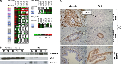
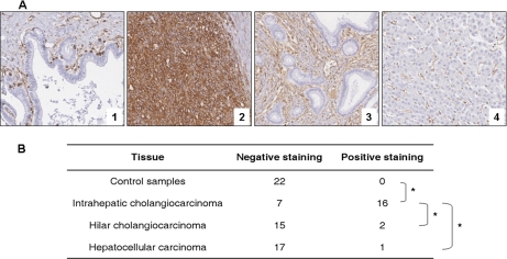
Obtaining accurate protein profiles from homogeneous cell populations in heterogeneous tissues can enhance the capability to discover protein biomarkers. In this context, methodologies to access specific cellular populations and analyze their proteome with exquisite sensitivity have to be selected. We report here the results of an investigation using a combination of laser microdissection and accurate mass and time tag proteomics. The study was aimed at the precise determination of proteome alterations in intrahepatic cholangiocarcinoma ICC, a markedly heterogeneous tumor. This cancer, which is difficult to diagnose and carries a very poor prognosis, has shown an unexplained increase in incidence over the last few years. Among a pool of 574 identified proteins, we were able to report on altered abundance patterns affecting 39 proteins conforming to a variety of potential tumorigenic pathways. The reliability of the proteomics results was confirmed by Western blot and immunohistochemistry on matched samples. Most of the proteins displaying perturbed abundances had not yet been described in the setting of ICC. These include proteins involved in cell mobility and actin cytoskeleton remodeling, which may participate in the epithelial to mesenchymal transition, a process invoked in migration and invasion of cancer cells. The biological relevance of these findings was explored using a tissue microarray. An increased abundance of vimentin was thus detected in 70% of ICC and none of the controls. These results suggest that vimentin could play a role in the aggressiveness of ICC and provide a basis for the serious outcome of this cancer.
Figures


Similar articles
-
Identification of TPD52 and DNAJB1 as two novel bile biomarkers for cholangiocarcinoma by iTRAQ‑based quantitative proteomics analysis.Oncol Rep. 2019 Dec;42(6):2622-2634. doi: 10.3892/or.2019.7387. Epub 2019 Oct 23. Oncol Rep. 2019. PMID: 31661142 Free PMC article.
-
A high level of integrin α6 expression in human intrahepatic cholangiocarcinoma cells is associated with a migratory and invasive phenotype.Dig Dis Sci. 2013 Jun;58(6):1627-35. doi: 10.1007/s10620-012-2524-6. Epub 2013 Jan 11. Dig Dis Sci. 2013. PMID: 23306848
-
FXR Acts as a Metastasis Suppressor in Intrahepatic Cholangiocarcinoma by Inhibiting IL-6-Induced Epithelial-Mesenchymal Transition.Cell Physiol Biochem. 2018;48(1):158-172. doi: 10.1159/000491715. Epub 2018 Jul 12. Cell Physiol Biochem. 2018. PMID: 30001540
-
Intrahepatic cholangiocarcinoma: new insights in pathology.Semin Liver Dis. 2011 Feb;31(1):49-60. doi: 10.1055/s-0031-1272839. Epub 2011 Feb 22. Semin Liver Dis. 2011. PMID: 21344350 Review.
-
The role of Tripartite motif containing 59 (TRIM59) in the proliferation and prognosis of intrahepatic cholangiocarcinoma.Pathol Res Pract. 2022 Aug;236:153989. doi: 10.1016/j.prp.2022.153989. Epub 2022 Jun 17. Pathol Res Pract. 2022. PMID: 35753134 Review.
Cited by
-
Ursodeoxycholic acid inhibits epithelial-mesenchymal transition, suppressing invasiveness of bile duct cancer cells: An in vitro study.Oncol Lett. 2022 Oct 26;24(6):448. doi: 10.3892/ol.2022.13568. eCollection 2022 Dec. Oncol Lett. 2022. PMID: 36420069 Free PMC article.
-
Beyond laser microdissection technology: follow the yellow brick road for cancer research.Am J Cancer Res. 2014 Jan 15;4(1):1-28. eCollection 2014. Am J Cancer Res. 2014. PMID: 24482735 Free PMC article. Review.
-
Differential Protein Expression Marks the Transition From Infection With Opisthorchis viverrini to Cholangiocarcinoma.Mol Cell Proteomics. 2017 May;16(5):911-923. doi: 10.1074/mcp.M116.064576. Epub 2017 Feb 23. Mol Cell Proteomics. 2017. PMID: 28232516 Free PMC article.
-
[Proteomics and translational medicine: molecular biomarkers for cancer diagnosis, prognosis and prediction of therapy outcome].Zhongguo Fei Ai Za Zhi. 2011 Aug;14(8):C6-9. doi: 10.3779/j.issn.1009-3419.2011.08.12. Zhongguo Fei Ai Za Zhi. 2011. PMID: 23676997 Free PMC article. Review. Chinese. No abstract available.
-
Identification of Four Immune Subtypes Characterized by Distinct Composition and Functions of Tumor Microenvironment in Intrahepatic Cholangiocarcinoma.Hepatology. 2020 Sep;72(3):965-981. doi: 10.1002/hep.31092. Epub 2020 Aug 16. Hepatology. 2020. PMID: 31875970 Free PMC article.
References
-
- Patel T. (2001) Increasing incidence and mortality of primary intrahepatic cholangiocarcinoma in the United States. Hepatology 33, 1353–1357 - PubMed
-
- Ben-Menachem T. (2007) Risk factors for cholangiocarcinoma. Eur. J. Gastroenterol. Hepatol. 19, 615–617 - PubMed
-
- Blechacz B. R., Gores G. J. (2008) Cholangiocarcinoma. Clin. Liver Dis. 12, 131–150, ix - PubMed
