Review
. 2010 Jul 7;102(13):932-41.
doi: 10.1093/jnci/djq187. Epub 2010 May 31.

Isocitrate dehydrogenase 1 and 2 mutations in cancer: alterations at a crossroads of cellular metabolism

Affiliations
Review

Isocitrate dehydrogenase 1 and 2 mutations in cancer: alterations at a crossroads of cellular metabolism

Zachary J Reitman et al. J Natl Cancer Inst. .

Abstract

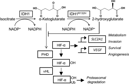
Dysregulation of metabolism is a common phenomenon in cancer cells. The NADP(+)-dependent isocitrate dehydrogenases 1 and 2 (IDH1 and IDH2) function at a crossroads of cellular metabolism in lipid synthesis, cellular defense against oxidative stress, oxidative respiration, and oxygen-sensing signal transduction. We review the normal functions of the encoded enzymes, frequent mutations of IDH1 and IDH2 recently found in human cancers, and possible roles for the mutated enzymes in human disease. IDH1 and IDH2 mutations occur frequently in some types of World Health Organization grades 2-4 gliomas and in acute myeloid leukemias with normal karyotype. IDH1 and IDH2 mutations are remarkably specific to codons that encode conserved functionally important arginines in the active site of each enzyme. To date, all IDH1 mutations have been identified at the Arg132 codon. Mutations in IDH2 have been identified at the Arg140 codon, as well as at Arg172, which is aligned with IDH1 Arg132. IDH1 and IDH2 mutations are usually heterozygous in cancer, and they appear to confer a neomorphic enzyme activity for the enzymes to catalyze the production of D-2-hydroxyglutarate. Study of alterations in these metabolic enzymes may provide insights into the metabolism of cancer cells and uncover novel avenues for development of anticancer therapeutics.


Figures

Figure 1
Figure 1
Isocitrate dehydrogenase 1 (IDH1) dimer in closed conformation. Structure of both molecules of the IDH1 dimer in the active closed conformation. The crystal structure of IDH1 is shown in ribbon format (PDBID:1T0L) (15). The dimer contains two active sites, each of which contains a NADP+-binding site and a metal ion–binding site. One active site is shown in the closed conformation, with the substrate isocitrate in dark blue and the cofactor NADP+ in red. Mutations that alter Arg132 (yellow) to histidine, cysteine, or other amino acids are associated with human gliomas and other cancers. This residue forms three hydrogen bonds with the isocitrate substrate (dark blue). Ser94 (orange) also forms one hydrogen bond with the isocitrate substrate. In the inactive open conformation (data not shown), Asp279 (cyan) contacts Ser94 and sterically hinders isocitrate binding. To transition to the active closed conformation shown here, Asp279 must swing away from Ser94 to relieve this steric hindrance. During this transition, Asp279 contacts Arg132 (15), suggesting that Arg132 plays a role in the transition between inactive and active enzyme conformations. Displayed image was created with University of California San Francisco Chimera software version 1.3 (San Francisco, CA) (16).
Figure 2
Figure 2
Functions of isocitrate dehydrogenase 1 (IDH1) and IDH2 in the normal cell. A) IDH1 and IDH2 catalyze the reversible conversion of isocitrate to α-ketoglutarate (α-KG) and NADP+ to NADPH. IDH1 is located in the cytosol and the peroxisome (–6). In the cytosol, IDH1 produces NADPH to contribute to the reduction of glutathione, the major cellular antioxidant (17). In the peroxisome, NADPH contributes to cholesterol synthesis (18). IDH1 has been shown to protect cells against gamma radiation (19), singlet oxygen (20), and UVB radiation (21) (light dashed line). IDH1 facilitates glucose-stimulated insulin secretion in pancreatic islets (22) (dashed arrows), which may be mediated by NADPH interactions with voltage-gated potassium channels or α-ketoglutarate interactions with α-ketoglutarate hydroxylases (data not shown). Conversion of α-ketoglutarate may be important for glia-specific glutamate and glutamine metabolism. IDH2 is located in the mitochondria and may function as the major catalyst of the isocitrate to α-ketoglutarate reaction in the tricarboxylic acid (TCA) cycle in some tissues (23). IDH3 is also located in the mitochondria and catalyzes the irreversible conversion of isocitrate to α-ketoglutarate and NAD+ to NADH. IDH2 has been shown to protect cells against gamma and ionizing radiation (19,24), high glucose (25), tumor necrosis factor-α (TNF-α) (26), and heat shock (27) (light dashed line). Carriers exchange malate (mal) for isocitrate, citrate, or α-ketoglutarate at the inner mitochondrial membrane. Cofactors for enzymes other than IDH1 and IDH2 are not indicated. B) A mitochondrial isocitrate/α-ketoglutarate cycle has been proposed by Sazanov and Jackson (28). In some tissues, flux through IDH2 proceeds in reverse, with α-ketoglutarate converted to isocitrate and NADPH converted to NADP+ (29). IDH3 converts isocitrate back to α-ketoglutarate and NAD+ to NADH. H+-transhydrogenase (H+-TH) completes this cycle by transferring electrons from NADH to NADPH (28). The transhydrogenase reaction is coupled to the transport of protons down their electrochemical potential gradient into the mitochondrial matrix (28). This cycle has the net effect of dissipating the electrochemical potential gradient across the inner mitochondrial membrane and producing heat. IDH2 also reduces the net flux from isocitrate to α-ketoglutarate (28). Because of this effect, allosteric modifiers of IDH3 (ATP, Ca2+, citrate, ATP, NADH, and NADPH) have a larger relative impact on net flux from isocitrate to α-ketoglutarate for the TCA cycle. DH = dehydrogenase; UVB = ultraviolet B.
Figure 3
Figure 3
Common genetic alterations in glioma tumorigenesis and progression. Grade 2 gliomas include well-differentiated oligodendrogliomas (O), oligoastrocytomas (OA), and diffuse astrocytomas (A) (72). All three types of tumors undergo two sequential genetic alterations for tumorigenesis: first, a mutation in isocitrate dehydrogenase 1 (IDH1) or IDH2, and second, homozygous deletion of chromosome arms 1p and 19q or tumor protein p53 (TP53) mutation. The IDH mutation event occurs at some point in the transformation from the glioma cell of origin to a glioma cell. The second genetic event contributes to the histopathological and clinical phenotype of the resulting tumor. For example, oligodendrogliomas usually contain 1p and 19q loss, whereas astrocytomas have TP53 mutations. The oligoastrocytomas can contain either genetic alteration. Grade 2 tumors can progress to grade 3 anaplastic oligodendrogliomas (AO), anaplastic oligoastrocytomas (AOA), and anaplastic astrocytomas (AA), as well as to grade 4 secondary glioblastomas (sGBM) and secondary glioblastomas with oligodendroglial component (sGBMO) (72). Grade 1 pilocytic astrocytomas (PA) and grade 4 primary GBMs (prGBM) arise de novo (72), do not frequently contain IDH mutations, and contain other alterations that are rare in the IDH mutation–containing tumors (54). It is not known whether these tumors arise from the same cell type. CDKN2A and 2B = cyclin-dependent kinase inhibitor 2A and 2B; EGFR = epidermal growth factor receptor; HD = homozygous deletion; PTEN = phosphatase and tensin homolog; WHO = World Health Organization.
Figure 4
Figure 4
Model for activation of hypoxia-inducible factor 1 (HIF-1) by isocitrate dehydrogenase 1 (IDH1) mutations. HIF-1α is hydroxylated by HIF prolyl hydroxylase (PHD), which targets HIF-1α for ubiquitylation by von Hippel Lindau protein (vHL) and subsequent proteasomal degradation. PHDs require O2 and α-ketoglutarate (α-KG) as substrates (82). When stabilized, HIF-1α dimerizes with HIF-1β and activates transcription of targets such as solute carrier family 2 member 1 (SLC2A1) and vascular endothelial growth factor (VEGF) (82). IDH1wt converts isocitrate to α-ketoglutarate, and IDH1R132H converts α-ketoglutarate to 2-hydroxyglutarate. By consuming α-ketoglutarate, IDH1R132H may lower the availability of this substrate, which would decrease PHD activity and lead to HIF-1α stabilization. Also, based on its structural similarity to α-ketoglutarate, 2-hydroxyglutarate has been hypothesized to competitively inhibit PHD activity by occupying PHD α-ketoglutarate binding sites (83). Ub = ubiquitin.

References

    1. Gabriel JL, Zervos PR, Plaut GW. Activity of purified NAD-specific isocitrate dehydrogenase at modulator and substrate concentrations approximating conditions in mitochondria. Metabolism. 1986;35(7):661–667. - PubMed
    1. Jennings GT, Sechi S, Stevenson PM, Tuckey RC, Parmelee D, McAlister-Henn L. Cytosolic NADP(+)-dependent isocitrate dehydrogenase. Isolation of rat cDNA and study of tissue-specific and developmental expression of mRNA. J Biol Chem. 1994;269(37):23128–23134. - PubMed
    1. Nekrutenko A, Hillis DM, Patton JC, Bradley RD, Baker RJ. Cytosolic isocitrate dehydrogenase in humans, mice, and voles and phylogenetic analysis of the enzyme family. Mol Biol Evol. 1998;15(12):1674–1684. - PubMed
    1. Geisbrecht BV, Gould SJ. The human PICD gene encodes a cytoplasmic and peroxisomal NADP(+)-dependent isocitrate dehydrogenase. J Biol Chem. 1999;274(43):30527–30533. - PubMed
    1. Henke B, Girzalsky W, Berteaux-Lecellier V, Erdmann R. IDP3 encodes a peroxisomal NADP-dependent isocitrate dehydrogenase required for the beta-oxidation of unsaturated fatty acids. J Biol Chem. 1998;273(6):3702–3711. - PubMed

Publication types

MeSH terms