. 2010 Sep;7(5):389-95.
doi: 10.1038/cmi.2010.28. Epub 2010 May 31.

Programmed death-1 upregulation is correlated with dysfunction of tumor-infiltrating CD8+ T lymphocytes in human non-small cell lung cancer

Affiliations

Programmed death-1 upregulation is correlated with dysfunction of tumor-infiltrating CD8+ T lymphocytes in human non-small cell lung cancer

Yan Zhang et al. Cell Mol Immunol. 2010 Sep.

Abstract

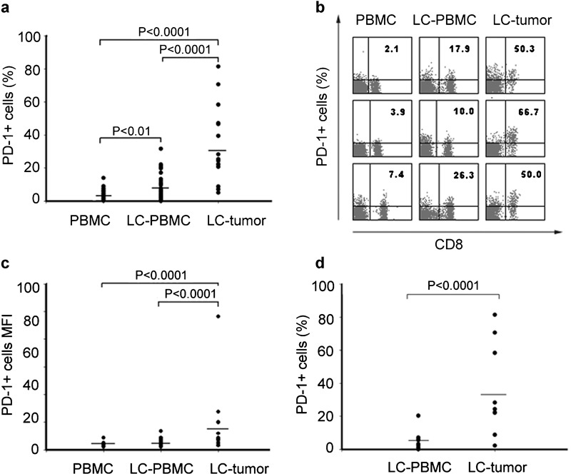
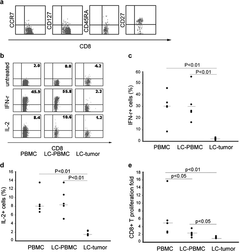
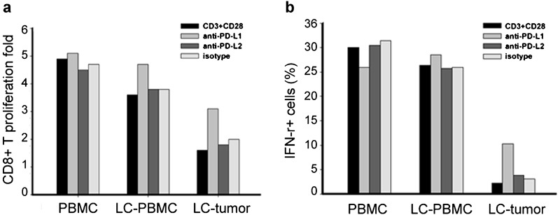
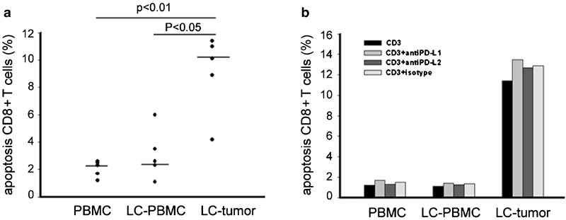
T-cell tolerance is an important mechanism for tumor escape, but the molecular pathways involved in T-cell tolerance remain poorly understood. It remains unknown whether the inhibitory immunoreceptor programmed death-1 (PD-1) plays a role in conditions of human non-small cell lung cancer (NSCLC). In this study, we detected PD-1 expression on CD8+ T cells from healthy control peripheral blood mononuclear cells (PBMCs) and the PBMCs of NSCLC patients as well as NSCLC tissues. Results showed that tumor-infiltrating CD8+ T cells had increased PD-1 expression and impaired immune function, including reducing cytokine production capability and impairing capacity to proliferate. Blockade of the PD-1/PD-L1 pathway by the PD-L1-specific antibody partially restored cytokine production and cell proliferation. These data provide direct evidence that the PD-1/PD-L1 pathway is involved in CD8+ T-cell dysfunction in NSCLC patients. Moreover, blocking this pathway provides a potential therapy target in lung cancer.


Figures

Figure 1
Figure 1
PD-1 is highly upregulated by tumor-infiltrating CD8+ T cells in patients with NSCLC. (a) Frequencies of PD-1-expressing CD8+ T cells in 58 blood samples, 16 lung tissue samples for LC patients and 23 blood samples for healthy controls. Each dot represents one individual. (b) PD-1 staining representative dot plots for one blood sample from a healthy person, as well as one blood sample and one tumor tissue sample from LC. Values in the upper right quadrant indicate the percentage of cells that express PD-1. (c) MFI of PD-1 expression on CD8+ T cells in PBMC and lung tissue samples for LC patients and healthy controls. Each dot represents one individual. (d) Positive rate of PD-1 in PBMCs and lung tissues from the same individual for 16 LC patients with. LC, lung cancer; MFI, mean fluorescence intensity; PBMC, peripheral blood mononuclear cell; PD-1, programmed death-1.
Figure 2
Figure 2
Preterminally differentiated phenotype and functional impairment of PD-1+ tumor-infiltrating CD8+ T cells. (a) Expression levels in PD-1+CD8+ T cells of CD45RA, CD127 (IL-7-receptor-α), CD27 and CCR7. (b) Representative dot plots of IFN-γ and IL-2 intracellular staining gated on CD8-positive cells for one blood sample from a healthy person, as well as one blood sample and one tumor tissue sample from LC. PBMCs and tumor tissues were stimulated with PMA and ionomycin for 6 h in the presence of 3 µg/ml of Brefeldin A and were stained with anti-CD8, fixed and permeabilized, followed by intracellular staining with antihuman IFN-γ or anti-IL-2 antibody. (c and d) Cytokine production in CD8+ T cells: IFN-γ (c) and IL-2 (d). (e) Proliferating fold of CD8-positive cells stimulated by anti-CD3 and anti-CD28. IFN, interferon; LC, lung cancer; PBMC, peripheral blood mononuclear cell; PD-1, programmed death-1; PMA, phorbol 12-myristate 13-acetate.
Figure 3
Figure 3
Blocking the PD-1/PD-L1 pathway increases production of effector molecules and proliferation of tumor-infiltrating CD8+ T cells. A purified CD8+ T-cell stimulation assay was performed in the presence of anti-CD3 and anti-CD28 alone and alternately with an antibody blockade of PD-L1 or PD-L2. (a) A fold of proliferating CD8-positive cells in the presence or absence of anti-PD-L1 or anti-PD-L2 antibodies. (b) Production of IFN-γ in the supernatants of 3-day cultures measured by ELISA in the presence or absence of anti-PD-L1 or anti-PD-L2. IFN, interferon; LC, lung cancer; PBMC, peripheral blood mononuclear cell; PD, programmed death.
Figure 4
Figure 4
Increased tumor-infiltrating CD8+ T-cell apoptosis and blocking the PD-1/PD-L1 pathway cannot abrogate apoptosis. (a) Increased apoptosis of tumor- infiltrating CD8+ T cells upon incubation with anti-CD3 for 3 days. Cells were harvested and examined for apoptosis by double staining with annexin V+ and an antibody against CD8. Apoptosis was calculated as the percentage of annexin V+ cells in CD8+ fraction. (b) Apoptosis of tumor-infiltrating CD8+ T cells in the presence of anti-PD-L1 or anti-PD-L2. LC, lung cancer; PBMC, peripheral blood mononuclear cell; PD, programmed death.

Comment in

References

    1. Jemal A, Siegel R, Ward E, Murray T, Xu J, Smigal C, et al. Cancer statistics, 2006. CA Cancer J Clin. 2006;56:106–130. - PubMed
    1. Lafferty KJ, Cunningham AJ. A new analysis of allogeneic interactions. Aust J Exp Biol Med Sci. 1975;53:27–42. - PubMed
    1. Swann JB, Smyth MJ. Immune surveillance of tumors. J Clin Invest. 2007;117:1137–1146. - PMC - PubMed
    1. Sharpe AH, Wherry EJ, Ahmed R, Freeman GJ. The function of programmed cell death 1 and its ligands in regulating autoimmunity and infection. Nat Immunol. 2007;8:239–245. - PubMed
    1. Barber DL, Wherry EJ, Masopust D, Zhu B, Allison JP, Sharpe AH, et al. Restoring function in exhausted CD8 T cells during chronic viral infection. Nature. 2006;439:682–687. - PubMed

Publication types

MeSH terms