

A new aspect of the TrkB signaling pathway in neural plasticity
- PMID: 20514207
- PMCID: PMC2811861
- DOI: 10.2174/157015909790031210
A new aspect of the TrkB signaling pathway in neural plasticity
Abstract
In the central nervous system (CNS), the expression of molecules is strictly regulated during development. Control of the spatiotemporal expression of molecules is a mechanism not only to construct the functional neuronal network but also to adjust the network in response to new information from outside of the individual, i.e., through learning and memory. Among the functional molecules in the CNS, one of the best-studied groups is the neurotrophins, which are nerve growth factor (NGF)-related gene family molecules. Neurotrophins include NGF, brain-derived neurotrophic factor (BDNF), neurotrophin 3 (NT-3), and NT-4/5 in the mammal. Among neurotrophins and their receptors, BDNF and tropomyosin-related kinases B (TrkB) are enriched in the CNS. In the CNS, the BDNF-TrkB signaling pathway fulfills a wide variety of functions throughout life, such as cell survival, migration, outgrowth of axons and dendrites, synaptogenesis, synaptic transmission, and remodeling of synapses. Although the same ligand and receptor, BDNF and TrkB, act in these various developmental events, we do not yet understand what kind of mechanism provokes the functional multiplicity of the BDNF-TrkB signaling pathway. In this review, we discuss the mechanism that elicits the variety of functions performed by the BDNF-TrkB signaling pathway in the CNS as a tool of pharmacological therapy.
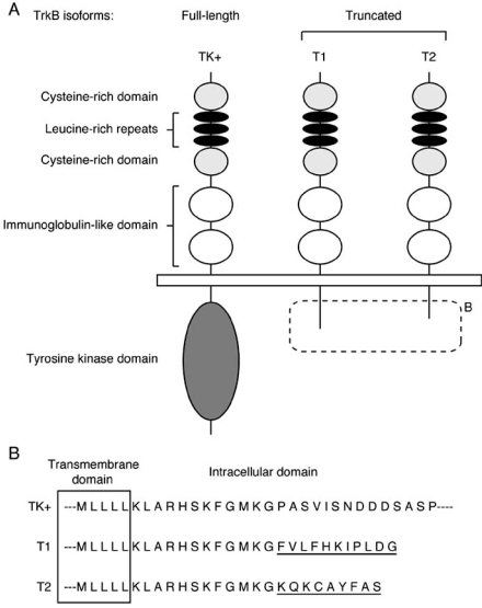
Keywords: Brain-derived neurotrophic factor; development; intracellular signaling; morphology; neural plasticity; neuron-glia interaction; receptor dimerization; truncated TrkB-T1..
Figures
References
-
- Aguado F, Carmona MA, Pozas E, Aguiló A, Matrínes-Guijarro FJ, Alcantara S, Borrell V, Yuste R, Ibañes CF, Soriano E. BDNF Regulates Spontaneous Correlated Activity at Early Developmental Stages by Increasing Synaptogenesis and Expression of the K+/Cl- Co-transporter KCC2. Development. 2003;130:1267–1280. - PubMed
-
- Alsina B, Vu T, Cohen-Cory S. Visualizing Synapse Formation in Arborizing Optic Axons in vivo: Dynamics and Modulation by BDNF. Nat. Neurosci. 2001;4:1093–1101. - PubMed
-
- Alter CA, Cai N, Bliven T, Juhansz M, Conner JM, Acheson AL, Lindsay RM, Wiegand SJ. Anterograde Transport of Brain-derived Neurotrophic Factor and Its Role in the Brain. Nature. 1997;389:856–860. - PubMed
LinkOut - more resources
Full Text Sources
Research Materials