

Case Reports
doi: 10.3346/jkms.2010.25.6.961.
Epub 2010 May 24.
A case of hypoglycemic brain injuries with cortical laminar necrosis
Affiliations
- PMID: 20514323
- PMCID: PMC2877241
- DOI: 10.3346/jkms.2010.25.6.961
Item in Clipboard
Case Reports
A case of hypoglycemic brain injuries with cortical laminar necrosis
J Korean Med Sci.
2010 Jun.
Abstract
We report a case of 68-yr-old male who died from brain injuries following an episode of prolonged hypoglycemia. While exploring controversies surrounding magnetic resonance imaging (MRI) findings indicating the bad prognosis in patients with hypoglycemia-induced brain injuries, we here discuss interesting diffusion-MRI of hypoglycemic brain injuries and their prognostic importance focusing on laminar necrosis of the cerebral cortex.
Keywords: Brain Injuries; Cerebral Cortical Necrosis; Diabetes; Diffusion Magnetic Resonance Imaging; Hypoglycemia; Prognosis.
Figures
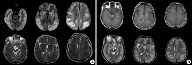
The initial magnetic resonance imaging. (A) Diffusion-Weighted MRI of the brain showed multiple bilateral hyperintense signals along the cortical and subcortical regions (frontal, temporal, parietal, and occipital lobes), hippocampus, caudate, globus pallidus, and putament and ADC (afferent diffusion coefficient) map showed low signal at the same area. (B) Low signal intensity lesion at T1-weighted image and High signal intensity lesion at T2 weighted image were seen at same area (Fig. 1B).
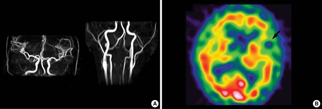
Angiography and single photon emission computed tomography (SPECT). (A) MR angiography of the brain and neck showed no abnormalities. (B) The SPECT with 99mTc-HMPAO showed focal hypoperfusion in the left temporal lobe.
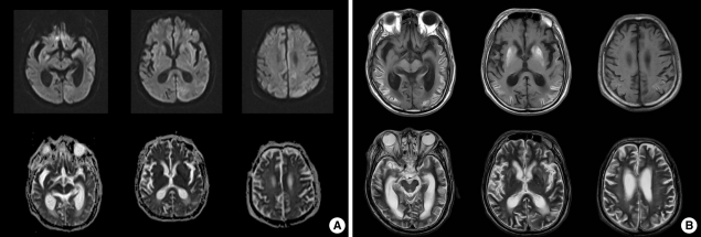
The follow-up magnetic resonance imaging. (A) Follow-up images on the 20th day show revisal of the hyperintensity lesions on diffusion-MRI and hypointensity lesion on ADC map curvilinear. (B) The T1 and T2-weighted image showed linear high signal intensity selectively along the cortical regions of bilateral hemisphere and basal ganglia.
Similar articles
-
Cortical laminar necrosis due to hypoglycaemic encephalopathy:-images in medicine.BMJ Case Rep. 2013 Feb 15;2013:bcr2012007726. doi: 10.1136/bcr-2012-007726. BMJ Case Rep. 2013. PMID: 23417931 Free PMC article. No abstract available.
-
Cerebral cortical laminar necrosis on diffusion-weighted MRI in hypoglycaemic encephalopathy.Diabet Med. 2005 Aug;22(8):1098-100. doi: 10.1111/j.1464-5491.2005.01568.x. Diabet Med. 2005. PMID: 16026379
-
[Clinical evaluation by MRI on the newborn infants with hypoglycemic brain damage].Zhonghua Er Ke Za Zhi. 2007 Jul;45(7):518-22. Zhonghua Er Ke Za Zhi. 2007. PMID: 17953809 Chinese.
-
Hypoglycemic brain injury.Semin Neonatol. 2001 Apr;6(2):147-55. doi: 10.1053/siny.2001.0044. Semin Neonatol. 2001. PMID: 11483020 Review.
-
MRI findings of hypoxic cortical laminar necrosis in a child with hemolytic anemia crisis.Eur Radiol. 2003 Dec;13 Suppl 6:L133-7. doi: 10.1007/s00330-002-1744-0. Epub 2002 Nov 22. Eur Radiol. 2003. PMID: 16440236 Review.
Cited by
-
Cortical laminar necrosis due to hypoglycaemic encephalopathy:-images in medicine.BMJ Case Rep. 2013 Feb 15;2013:bcr2012007726. doi: 10.1136/bcr-2012-007726. BMJ Case Rep. 2013. PMID: 23417931 Free PMC article. No abstract available.
-
Acute Cortical Lesions in MELAS Syndrome: Anatomic Distribution, Symmetry, and Evolution.AJNR Am J Neuroradiol. 2020 Jan;41(1):167-173. doi: 10.3174/ajnr.A6325. Epub 2019 Dec 5. AJNR Am J Neuroradiol. 2020. PMID: 31806591 Free PMC article.
-
Hypoglycemic encephalopathy: a case series and literature review on outcome determination.J Neurol. 2012 Oct;259(10):2172-81. doi: 10.1007/s00415-012-6480-z. Epub 2012 Apr 11. J Neurol. 2012. PMID: 22491856 Review.
-
Brain Injury in Neonatal Hypoglycemia: A Hospital-Based Cohort Study.Clin Med Insights Pediatr. 2019 Aug 8;13:1179556519867953. doi: 10.1177/1179556519867953. eCollection 2019. Clin Med Insights Pediatr. 2019. PMID: 31447599 Free PMC article.
-
The radiological findings of hypoglycemic encephalopathy: A case report with high b value DWI analysis.Medicine (Baltimore). 2017 Oct;96(43):e8425. doi: 10.1097/MD.0000000000008425. Medicine (Baltimore). 2017. PMID: 29069042 Free PMC article.
References
-
- Cho SJ, Minn YK, Kwon KH. Severe hypoglycemia and vulnerability of the brain. Arch Neurol. 2006;63:138. - PubMed
-
- Aoki T, Sato T, Hasegawa K, Ishizaki R, Saiki M. Reversible hyperintensity lesion on diffusion-weighted MRI in hypoglycemic coma. Neurology. 2004;63:392–393. - PubMed
-
- Anderson JM, Milner RD, Strich SJ. Pathological changes in the nervous system in severe neonatal hypoglycaemia. Lancet. 1966;2:372–375. - PubMed
-
- Yoneda Y, Yamamoto S. Cerebral cortical laminar necrosis on diffusion-weighted MRI in hypoglycaemic encephalopathy. Diabet Med. 2005;22:1098–1100. - PubMed
-
- Chan R, Erbay S, Oljeski S, Thaler D, Bhadelia R. Case report: hypoglycemia and diffusion-weighted imaging. J Comput Assist Tomogr. 2003;27:420–423. - PubMed
Publication types
MeSH terms
LinkOut - more resources
Full Text Sources
Medical
Research Materials
