BMPs in periprosthetic tissues around aseptically loosened total hip implants
- PMID: 20515435
- PMCID: PMC2917563
- DOI: 10.3109/17453674.2010.492765
BMPs in periprosthetic tissues around aseptically loosened total hip implants
Abstract
Background and purpose: Primary and dynamically maintained periprosthetic bone formation is essential for osseointegration of hip implants to host bone. Bone morphogenetic proteins (BMPs) play a role in osteoinductive bone formation. We hypothesized that there is an increased local synthesis of BMPs in the synovial membrane-like interface around aseptically loosened total hip replacement (THR) implants, as body attempts to generate or maintain implant fixation.
Patients and methods: We compared synovial membrane-like interface tissue from revised total hip replacements (rTHR, n = 9) to osteoarthritic control synovial membrane samples (OA, n = 11. Avidin-biotin-peroxidase complex staining and grading of BMP-2, BMP-4, BMP-6, and BMP-7 was done. Immunofluorescence staining was used to study BMP proteins produced by mesenchymal stromal/stem cells (MSCs) and osteoblasts.
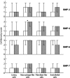
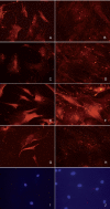
Results and interpretation: All BMPs studied were present in the synovial lining or lining-like layer, fibroblast-like stromal cells, interstitial macrophage-like cells, and endothelial cells. In OA and rTHR samples, BMP-6 positivity in cells, inducible by the proinflammatory cytokines tumor necrosis factor-alpha and interleukin-1beta, predominated over expression of other BMPs. Macrophage-like cells positive for BMP-4, inducible in macrophages by stimulation with particles, were more frequent around loosened implants than in control OA samples, but apparently not enough to prevent loosening. MSCs contained BMP-2, BMP-4, BMP-6, and BMP-7, but this staining diminished during osteogenesis, suggesting that BMPs are produced by progenitor cells in particular, probably for storage in the bone matrix.
Figures


Similar articles
-
Production of platelet-derived growth factor in aseptic loosening of total hip replacement.Rheumatol Int. 1998;17(6):215-21. doi: 10.1007/s002960050037. Rheumatol Int. 1998. PMID: 9592860
-
Role and regulation of VEGF and its receptors 1 and 2 in the aseptic loosening of total hip implants.J Orthop Res. 2012 Nov;30(11):1830-6. doi: 10.1002/jor.22138. Epub 2012 Apr 23. J Orthop Res. 2012. PMID: 22528855
-
Expression of parathyroid hormone related protein in the tissue around loosened hip prostheses.Clin Exp Rheumatol. 2001 Nov-Dec;19(6):689-95. Clin Exp Rheumatol. 2001. PMID: 11791641
-
Bone morphogenetic proteins.Growth Factors. 2004 Dec;22(4):233-41. doi: 10.1080/08977190412331279890. Growth Factors. 2004. PMID: 15621726 Review.
-
Current methods of preventing aseptic loosening and improving osseointegration of titanium implants in cementless total hip arthroplasty: a review.J Int Med Res. 2018 Jun;46(6):2104-2119. doi: 10.1177/0300060517732697. Epub 2017 Nov 3. J Int Med Res. 2018. PMID: 29098919 Free PMC article. Review.
Cited by
-
VDR Gene Polymorphisms (BsmI, FokI, TaqI, ApaI) in Total Hip Arthroplasty Outcome Patients.Int J Mol Sci. 2024 Jul 27;25(15):8225. doi: 10.3390/ijms25158225. Int J Mol Sci. 2024. PMID: 39125794 Free PMC article.
-
Peri-prosthetic tissue cells show osteogenic capacity to differentiate into the osteoblastic lineage.J Orthop Res. 2017 Aug;35(8):1732-1742. doi: 10.1002/jor.23457. Epub 2017 Apr 13. J Orthop Res. 2017. PMID: 27714894 Free PMC article.
References
-
- Anderson HC, Hodges PT, Aguilera XM, Missana L, Moylan PE. Bone morphogenetic protein (BMP) localization in developing human and rat growth plate, metaphysis, epiphysis, and articular cartilage. J Histochem Cytochem. 2000;48:1493–502. - PubMed
-
- Badlani N, Inoue A, Healey R, Coutts R, Amiel D. The protective effect of OP-1 on articular cartilage in the development of osteoarthritis. Osteoarthritis Cartilage. 2008;16:600–6. - PubMed
-
- Blom AB, van Lent PL, Holthuysen AE, van der Kraan PM, Roth J, van Rooijen N, van den Berg WB. Synovial lining macrophages mediate osteophyte formation during experimental osteoarthritis. Osteoarthritis Cartilage. 2004;12:627–35. - PubMed
-
- Bragdon CR, Doherty AM, Rubash HE, Jasty M, Li XJ, Seeherman H, Harris WH. The efficacy of BMP-2 to induce bone ingrowth in a total hip replacement model. Clin Orthop. 2003;((417)):50–61. - PubMed
Publication types
MeSH terms
Substances
LinkOut - more resources
Full Text Sources
Medical
