. 2010 Aug 1;429(3):473-83.
doi: 10.1042/BJ20100155.

The potency of the fs260 connexin43 mutant to impair keratinocyte differentiation is distinct from other disease-linked connexin43 mutants

Affiliations

The potency of the fs260 connexin43 mutant to impair keratinocyte differentiation is distinct from other disease-linked connexin43 mutants

Jared M Churko et al. Biochem J. .

Abstract

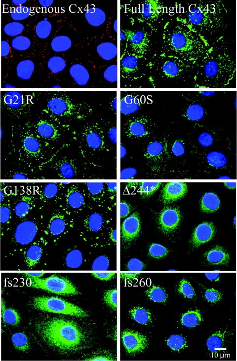
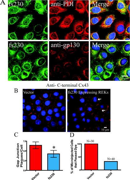
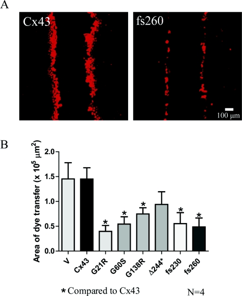
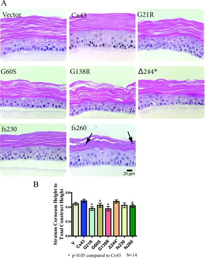
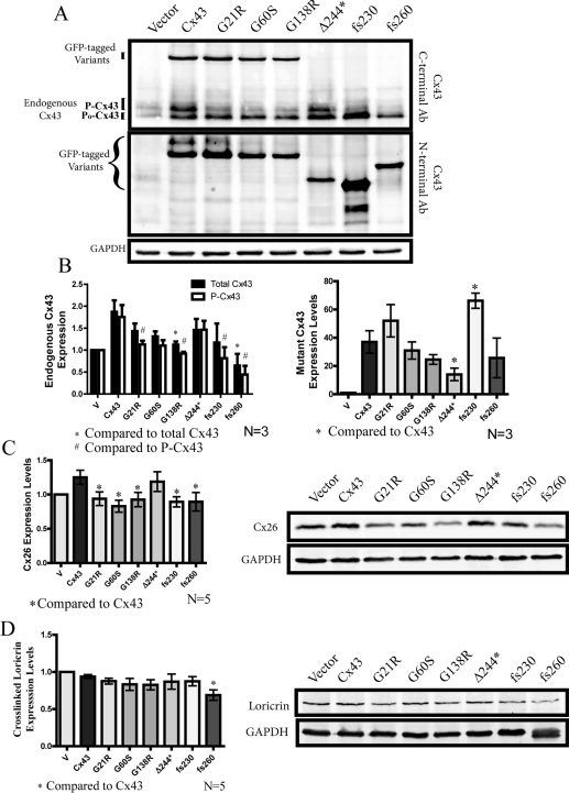
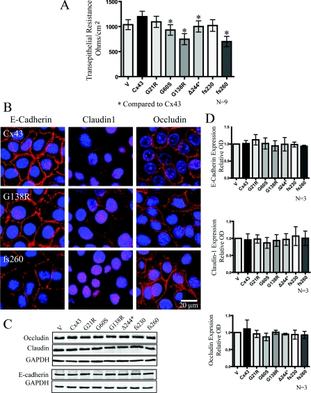
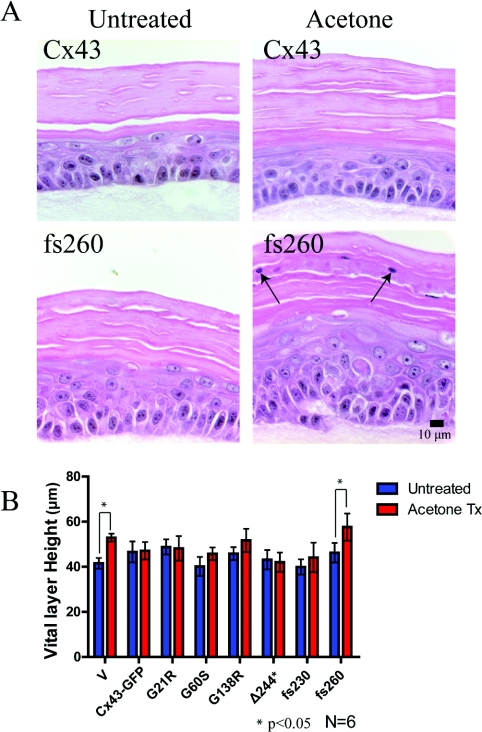
Although there are currently 62 mutants of Cx43 (connexin43) that can cause ODDD (oculodentodigital dysplasia), only two mutants have also been reported to cause palmar plantar hyperkeratosis. To determine how mutants of Cx43 can lead to this skin disease, REKs (rat epidermal keratinocytes) were engineered to express an ODDD-associated Cx43 mutant always linked to skin disease (fs260), an ODDD-linked Cx43 mutant which has been reported to sometimes cause skin disease (fs230), Cx43 mutants which cause ODDD only (G21R, G138R), a mouse Cx43 mutant linked to ODDD (G60S), a non-disease-linked truncated Cx43 mutant that is trapped in the endoplasmic reticulum (Delta244*) or full-length Cx43. When grown in organotypic cultures, of all the mutants investigated, only the fs260-expressing REKs consistently developed a thinner stratum corneum and expressed lower levels of Cx43, Cx26 and loricrin in comparison with REKs overexpressing wild-type Cx43. REKs expressing the fs260 mutant also developed a larger organotypic vital layer after acetone-induced injury and exhibited characteristics of parakeratosis. Collectively, our results suggest that the increased skin disease burden exhibited in ODDD patients harbouring the fs260 mutant is probably due to multiple additive effects cause by the mutant during epidermal differentiation.


Figures

Figure 1
Figure 1. Cx43 mutants display different localization profiles in REKs
REKs were engineered by retroviral infection to stably express GFP-tagged full-length Cx43 (Cx43), two ODDD mutants that cause skin disease (fs230 and fs260), a C-terminal truncation with two additional mutations (Δ244*), as well as three ODDD-linked mutants not associated with skin disease (G21R, G60S and G138R). Full-length Cx43, as well as the G21R and G138R mutants, traffic to the plasma membrane and display large gap junctions, whereas G60S, Δ244*, fs230 and fs260 mutant expression is largely confined to intracellular compartments. Endogenous Cx43 was detected by immunolabelling (red) and nuclei were labelled with DAPI (blue).
Figure 2
Figure 2. The fs230 mutant is predominantly localized within the endoplasmic reticulum and reduces the number of endogenous gap junctions and the extent of GJIC
(A) fs230-expressing REKs were labelled with antibodies specific for PDI (endoplasmic reticulum-resident protein) or gp130 (Golgi marker). The bulk of fs230 co-localized primarily with PDI. Nuclei were stained with DAPI (blue). (B) Both vector control and fs230-expressing REKs were immunolabelled with a C-terminal specific anti-Cx43 antibody that detected only endogenous Cx43. Arrows indicate plaque formation. (C) fs230-expressing REKs exhibited a 34% decrease in endogenous Cx43 plaque formation when compared with vector control REKs. (D) Lucifer Yellow dye was microinjected into both vector control and fs230-expressing REKs. All vector control REKs passed Lucifer Yellow dye, whereas only 33% of fs230-expressing REKs passed dye to neighbouring cells. *P<0.05.
Figure 3
Figure 3. Cx43 mutants reduce dye transfer in REKs
(A) To assess the ability of REKs to transfer dye to neighbouring cells, a scrape assay was performed using a gap junction permeable dye, sulforhodamine. (B) With the exception of Δ244*, there was a significant reduction in dye spread in mutant expressing REKs compared with Cx43-overexpressing REKs. V, empty vector. *P<0.05.
Figure 4
Figure 4. Cx43 mutant-expressing REKs differentiate into organotypic epidermis
REKs were plated on to collagen type I-coated well inserts and the medium from the upper chamber was removed to allow REKs to differentiate and stratify. (A) After two weeks of organotypic differentiation, all mutant expressing Cx43 REK cell lines differentiated. To determine the extent of organotypic differentiation, histological sections of each organotypic culture were stained with haematoxylin and eosin, imaged, and the upper stratum corneum and lower vital layer thicknesses were measured. (B) When comparing the stratum corneum height with total epidermal height, there was a significant decrease found in the G21R-, G60S-, G138R- and fs260-expressing cultures when compared with organotypic cultures derived from Cx43 expressing REKs. In addition, parakeratosis was observed in the fs260-expressing cultures (arrows). *P<0.05.
Figure 5
Figure 5. Assessment of endogenous and mutant Cx43, Cx26 and loricrin protein expression in organotypic epidermis
Western blots were performed on REK cells lines after 2 weeks of differentiation. (A) Anti-Cx43 N-terminal-specific antibody (Ab) was used to detect GFP-tagged mutant Cx43 variants, whereas an anti-Cx43 C-terminal-specific antibody was used to detect endogenous Cx43 expression. (B) The G138R- and fs260-expressing REKs had significantly lower total endogenous Cx43 levels when compared with full-length Cx43 REKs. ODDD mutants also reduced the highly phosphorylated species of endogenous Cx43. Densitometry analysis of GFP-tagged Cx43 variants also revealed a significant decrease in the expression of the Δ244* mutant, whereas the expression of the fs230 mutant was signficantly elevated. In addition, Western blot analysis of Cx26 (C) revealed that all ODDD Cx43 mutants reduced the levels of Cx26 compared with cells expressing full-length Cx43, whereas loricrin (D) was only reduced in fs260-expressing REKs. *P<0.05 and #P<0.05.
Figure 6
Figure 6. Cx43 mutant-expressing REKs exhibited reduced transepithelial resistance when compared with Cx43 overexpressing REKs in the absence of changes in the expression or localization of junctional proteins
(A) Transepithelial resistance measurements of confluent monolayer cultures were taken daily for 5 days and the resistance from days 2–4 were averaged and plotted. (B) E-cadherin, claudin1 and occludin were localized by immunofluorescent labelling in REKs overexpressing GFP-tagged Cx43, G138R or fs260. Western blot analysis did not reveal any changes in the expression of occludin, E-cadherin or claudin-1 (C and D). *P<0.05. Nuclei were labelled with DAPI (blue). GAPDH, glyceraldehyde-3-phosphate dehydrogenase
Figure 7
Figure 7. Acetone treatment increased the vital layer thickness in fs260-expressing organotypic cultures
(A) Organotypic cultures of REKs expressing endogenous Cx43 or overexpressing Cx43 or Cx43 mutants were left untreated or treated with acetone. Note that in fs260-expressing REK epidermis, there were nuclei present in the stratum corneum (arrows). (B) Vital layer measurements of all REK cell lines either untreated or treated with acetone revealed a significant increase in the vital layer thickness in both the vector and fs260-expressing REKs when compared with full-length Cx43 expressing organotypic cultures. *P<0.05.

Similar articles

Cited by

References

    1. Beyer E. C., Paul D. L., Goodenough D. A. Connexin43: a protein from rat heart homologous to a gap junction protein from liver. J. Cell Biol. 1987;105:2621–2629. - PMC - PubMed
    1. Vanslyke J. K., Naus C. C., Musil L. S. Conformational maturation and post-ER multisubunit assembly of gap junction proteins. Mol. Biol. Cell. 2009;20:2451–2463. - PMC - PubMed
    1. Goodenough D. A., Goliger J. A., Paul D. L. Connexins, connexons, and intercellular communication. Annu. Rev. Biochem. 1996;65:475–502. - PubMed
    1. Gong X. Q., Shao Q., Lounsbury C. S., Bai D., Laird D. W. Functional characterization of a GJA1 frameshift mutation causing oculodentodigital dysplasia and palmoplantar keratoderma. J. Biol. Chem. 2006;281:31801–31811. - PubMed
    1. Lai A., Le D. N., Paznekas W. A., Gifford W. D., Jabs E. W., Charles A. C. Oculodentodigital dysplasia connexin43 mutations result in non-functional connexin hemichannels and gap junctions in C6 glioma cells. J. Cell Sci. 2006;119:532–541. - PubMed

Publication types