

Equivalent moving dipole localization of cardiac ectopic activity in a swine model during pacing
- PMID: 20515710
- PMCID: PMC2948591
- DOI: 10.1109/TITB.2010.2051448
Equivalent moving dipole localization of cardiac ectopic activity in a swine model during pacing
Abstract
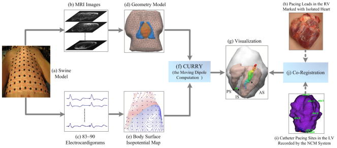
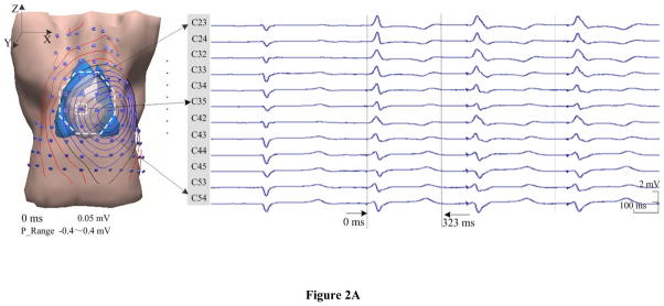
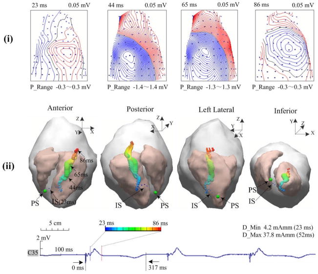
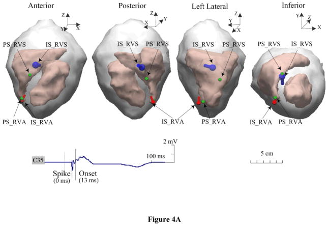
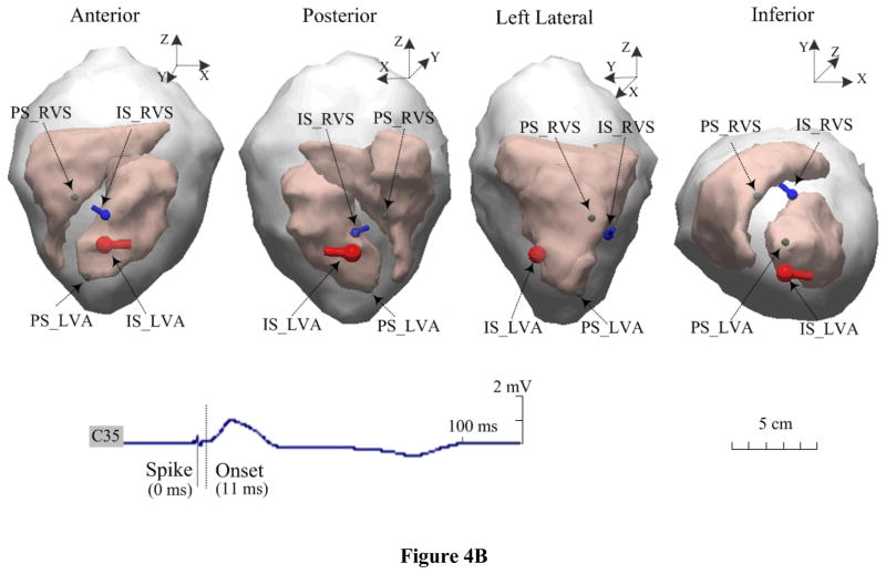
Localization of the initial site of cardiac ectopic activity has direct clinical benefits for treating focal cardiac arrhythmias. The aim of the present study is to experimentally evaluate the performance of the equivalent moving dipole technique on noninvasively localizing the origin of the cardiac ectopic activity from the recorded body surface potential mapping (BSPM) data in a well-controlled experimental setting. The cardiac ectopic activities were induced in four well-controlled intact pigs by either single-site pacing or dual-site pacing within the ventricles. In each pacing study, the initiation sites of cardiac ectopic activity were localized by estimating the locations of a single moving dipole (SMD) or two moving dipoles (TMDs) from the measured BSPM data and compared with the precise pacing sites (PSs). For the single-site pacing, the averaged SMD localization error was 18.6 ± 3.8 mm over 16 sites, while the averaged distance between the TMD locations and the two corresponding PSs was slightly larger (24.9 ± 6.2 mm over five pairs of sites), both occurring at the onset of the QRS complex (10-25 ms following the pacing spike). The obtained SMD trajectories originated near the stimulus site and then traversed across the heart during the ventricular depolarization. The present experimental results show that the initial location of the moving dipole can provide the approximate site of origin of a cardiac ectopic activity in vivo, and that the migration of the dipole can portray the passage of an ectopic beat across the heart.
Figures

Similar articles
-
Cardiac source localization by means of a single moving dipole solution during endocardial pacing in an animal model.Annu Int Conf IEEE Eng Med Biol Soc. 2009;2009:1778-80. doi: 10.1109/IEMBS.2009.5334014. Annu Int Conf IEEE Eng Med Biol Soc. 2009. PMID: 19964556
-
Localization of endocardial ectopic activity by means of noninvasive endocardial surface current density reconstruction.Phys Med Biol. 2011 Jul 7;56(13):4161-76. doi: 10.1088/0031-9155/56/13/027. Epub 2011 Jun 21. Phys Med Biol. 2011. PMID: 21693786 Free PMC article.
-
Localization of cardiac ectopic activity in man by a single moving dipole. Comparison of different computation techniques.J Electrocardiol. 1985 Jul;18(3):211-21. doi: 10.1016/s0022-0736(85)80045-5. J Electrocardiol. 1985. PMID: 4031724
-
Rapid 12-lead automated localization method: Comparison to electrocardiographic imaging (ECGI) in patient-specific geometry.J Electrocardiol. 2018 Nov-Dec;51(6S):S92-S97. doi: 10.1016/j.jelectrocard.2018.07.022. Epub 2018 Jul 29. J Electrocardiol. 2018. PMID: 30177365
-
The single equivalent moving dipole model does not require spatial anatomical information to determine cardiac sources of activation.IEEE J Biomed Health Inform. 2014 Jan;18(1):222-30. doi: 10.1109/JBHI.2013.2268012. IEEE J Biomed Health Inform. 2014. PMID: 24403420
Cited by
-
Noninvasive Activation Imaging of Ventricular Arrhythmias by Spatial Gradient Sparse in Frequency Domain-Application to Mapping Reentrant Ventricular Tachycardia.IEEE Trans Med Imaging. 2019 Feb;38(2):525-539. doi: 10.1109/TMI.2018.2866951. Epub 2018 Aug 23. IEEE Trans Med Imaging. 2019. PMID: 30136937 Free PMC article.
-
Simulation-based validation for four- dimensional multi-channel ultrasound current source density imaging.IEEE Trans Ultrason Ferroelectr Freq Control. 2014 Mar;61(3):420-7. doi: 10.1109/TUFFC.2014.2927. IEEE Trans Ultrason Ferroelectr Freq Control. 2014. PMID: 24569247 Free PMC article.
-
Imaging cardiac activation sequence during ventricular tachycardia in a canine model of nonischemic heart failure.Am J Physiol Heart Circ Physiol. 2015 Jan 15;308(2):H108-14. doi: 10.1152/ajpheart.00196.2014. Epub 2014 Nov 21. Am J Physiol Heart Circ Physiol. 2015. PMID: 25416188 Free PMC article.
-
Usefulness of ventricular endocardial electric reconstruction from body surface potential maps to noninvasively localize ventricular ectopic activity in patients.Phys Med Biol. 2013 Jun 7;58(11):3897-909. doi: 10.1088/0031-9155/58/11/3897. Epub 2013 May 16. Phys Med Biol. 2013. PMID: 23681281 Free PMC article.
-
Noninvasive imaging of 3-dimensional myocardial infarction from the inverse solution of equivalent current density in pathological hearts.IEEE Trans Biomed Eng. 2015 Feb;62(2):468-76. doi: 10.1109/TBME.2014.2358618. Epub 2014 Sep 17. IEEE Trans Biomed Eng. 2015. PMID: 25248174 Free PMC article.
References
-
- Gulrajani RM, Roberge FA, Savard P. Moving dipole inverse ECG and EEG solutions. IEEE Trans Biomed Eng. 1984 Dec;31(12):903–910. - PubMed
-
- He B, Wu D. Imaging and visualization of 3D cardiac electric activity. IEEE Trans Inf Technol Biomed. 2001 Sept;5(3):181–186. - PubMed
-
- Li G, He B. Localization of the site of origin of cardiac activation by means of heart-model-based electrocardiographic imaging approach. IEEE Trans Biomed Eng. 2001 Jun;48(6):660–669. - PubMed
-
- He B, Li G, Zhang X. Noninvasive three-dimensional activation time imaging of ventricular excitation by means of a heart-excitation-model. Phys Med Biol. 2002 Nov;47:4063–4078. - PubMed
