

Identification and characterization of the promoter of fibroblast activation protein
- PMID: 20515787
- PMCID: PMC2880831
- DOI: 10.2741/e175
Identification and characterization of the promoter of fibroblast activation protein
Abstract
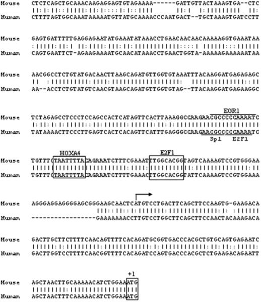
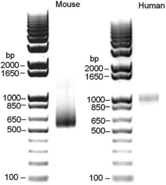
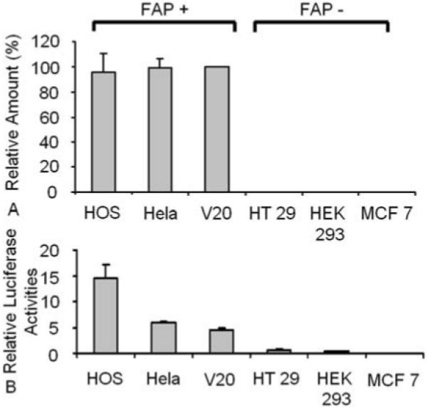
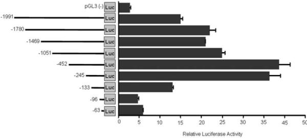
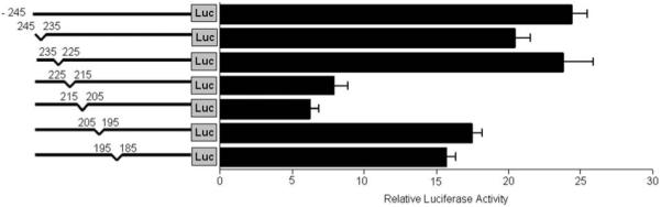
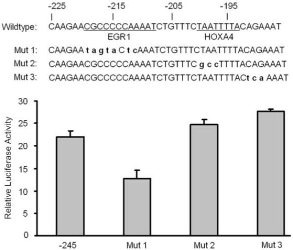
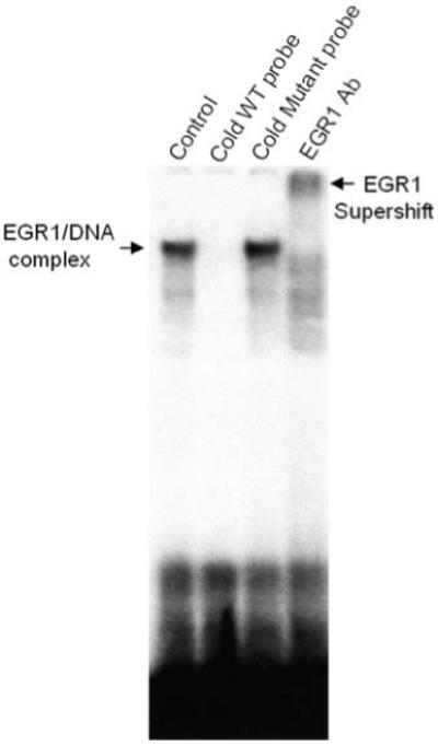
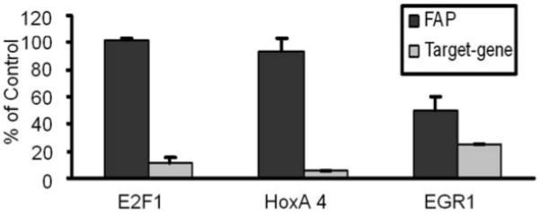
Fibroblast activation protein (FAP) is a type II integral membrane glycoprotein belonging to the serine protease family. It is selectively expressed by tumor stromal fibroblasts and transiently in the fibroblasts of healing wounds. FAP has been shown to modulate growth, differentiation, adhesion, and metastasis of tumor cells. Despite the importance of FAP in cancer, the mechanisms that govern its expression have not been defined. In this study, we determined the transcription start site of the FAP gene and identified a 2-kb segment with promoter activity in cells expressing FAP. Truncation of this fragment revealed that the core promoter activity resided in a 245-bp fragment surrounding the transcription start site. Electrophoretic mobility shift assay showed that EGR1 binds to the FAP promoter. Mutation of the EGR1 site within this fragment significantly decreased the promoter activity of FAP and eliminated EGR1 binding. Down-regulation of EGR1 resulted in a significant reduction in endogenous FAP mRNA expression. These findings identify the basal transcriptional requirements of FAP gene expression and show EGR1 is an important regulator of FAP expression.
Figures
Similar articles
-
Molecular cloning of fibroblast activation protein alpha, a member of the serine protease family selectively expressed in stromal fibroblasts of epithelial cancers.Proc Natl Acad Sci U S A. 1994 Jun 7;91(12):5657-61. doi: 10.1073/pnas.91.12.5657. Proc Natl Acad Sci U S A. 1994. PMID: 7911242 Free PMC article.
-
Effects of the fibroblast activation protein on the invasion and migration of gastric cancer.Exp Mol Pathol. 2013 Dec;95(3):350-56. Exp Mol Pathol. 2013. PMID: 24422232
-
Mouse fibroblast-activation protein--conserved Fap gene organization and biochemical function as a serine protease.Eur J Biochem. 1998 Jun 15;254(3):650-4. doi: 10.1046/j.1432-1327.1998.2540650.x. Eur J Biochem. 1998. PMID: 9688278
-
The role of fibroblast activation protein in health and malignancy.Cancer Metastasis Rev. 2020 Sep;39(3):783-803. doi: 10.1007/s10555-020-09909-3. Cancer Metastasis Rev. 2020. PMID: 32601975 Free PMC article. Review.
-
Targeting fibroblast activation protein in cancer - Prospects and caveats.Front Biosci (Landmark Ed). 2018 Jun 1;23(10):1933-1968. doi: 10.2741/4682. Front Biosci (Landmark Ed). 2018. PMID: 29772538 Review.
Cited by
-
Targeting FAPα-expressing hepatic stellate cells overcomes resistance to antiangiogenics in colorectal cancer liver metastasis models.J Clin Invest. 2022 Oct 3;132(19):e157399. doi: 10.1172/JCI157399. J Clin Invest. 2022. PMID: 35951441 Free PMC article.
-
Tumors as organs: biologically augmenting radiation therapy by inhibiting transforming growth factor β activity in carcinomas.Semin Radiat Oncol. 2013 Oct;23(4):242-51. doi: 10.1016/j.semradonc.2013.05.001. Semin Radiat Oncol. 2013. PMID: 24012338 Free PMC article. Review.
-
Pro-tumorigenic roles of fibroblast activation protein in cancer: back to the basics.Oncogene. 2018 Aug;37(32):4343-4357. doi: 10.1038/s41388-018-0275-3. Epub 2018 May 3. Oncogene. 2018. PMID: 29720723 Free PMC article. Review.
-
The Role and Application of Fibroblast Activating Protein.Curr Mol Med. 2024;24(9):1097-1110. doi: 10.2174/1566524023666230530095305. Curr Mol Med. 2024. PMID: 37259211 Review.
-
The role of fibroblast activation protein in progression and development of osteosarcoma cells.Clin Exp Med. 2020 Feb;20(1):121-130. doi: 10.1007/s10238-019-00591-6. Epub 2019 Nov 19. Clin Exp Med. 2020. Retraction in: Clin Exp Med. 2023 Dec;23(8):5481. doi: 10.1007/s10238-023-01211-0. PMID: 31745677 Retracted.
References
-
- Rettig WJ, Chesa PG, Beresford HR, Feickert HJ, Jennings MT, Cohen J, Oettgen HF, Old LJ. Differential expression of cell surface antigens and glial fibrillary acidic protein in human astrocytoma subsets. Cancer Res. 1986;46(12 Pt 1):6406–12. - PubMed
-
- Niedermeyer J, Enenkel B, Park JE, Lenter M, Rettig WJ, Damm K, Schnapp A. Mouse fibroblast-activation protein--conserved Fap gene organization and biochemical function as a serine protease. Eur J Biochem. 1998;254(3):650–4. - PubMed
-
- Park JE, Lenter MC, Zimmermann RN, Garin-Chesa P, Old LJ, Rettig WJ. Fibroblast activation protein, a dual specificity serine protease expressed in reactive human tumor stromal fibroblasts. J Biol Chem. 1999;274(51):36505–12. - PubMed
-
- Mathew S, Scanlan MJ, Mohan Raj BK, Murty VV, Garin-Chesa P, Old LJ, Rettig WJ, Chaganti RS. The gene for fibroblast activation protein alpha (FAP), a putative cell surface-bound serine protease expressed in cancer stroma and wound healing, maps to chromosome band 2q23. Genomics. 1995;25(1):335–7. - PubMed
Publication types
MeSH terms
Substances
Grants and funding
LinkOut - more resources
Full Text Sources
Other Literature Sources
Miscellaneous
