. 2010 Aug;78(8):3465-74.
doi: 10.1128/IAI.00406-10. Epub 2010 Jun 1.

Coxiella burnetii phase I and II variants replicate with similar kinetics in degradative phagolysosome-like compartments of human macrophages

Affiliations

Coxiella burnetii phase I and II variants replicate with similar kinetics in degradative phagolysosome-like compartments of human macrophages

Dale Howe et al. Infect Immun. 2010 Aug.

Abstract

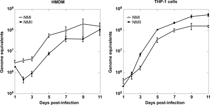
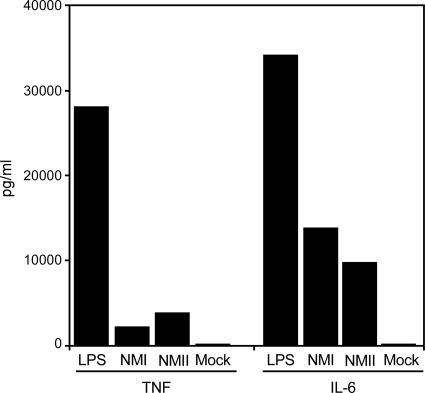
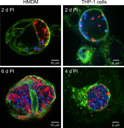
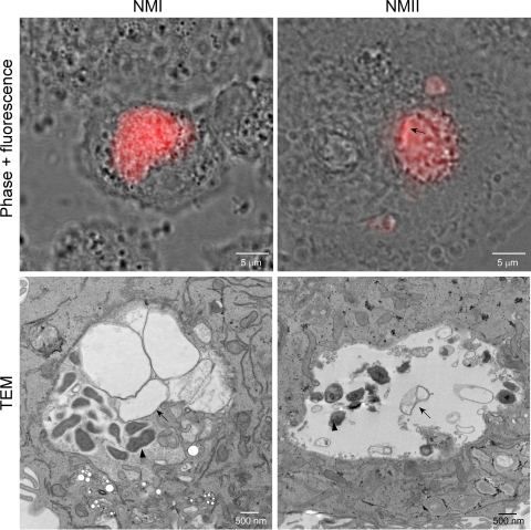
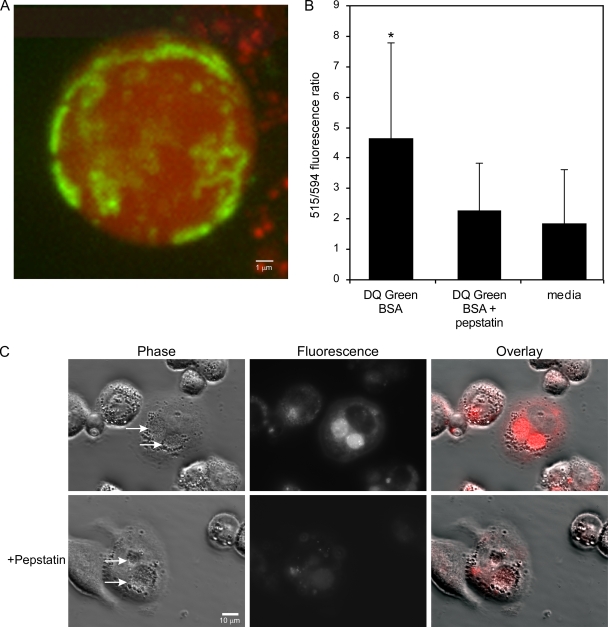
Coxiella burnetii infects mononuclear phagocytes, where it directs biogenesis of a vacuolar niche termed the parasitophorous vacuole (PV). Owing to its lumenal pH (approximately 5) and fusion with endolysosomal vesicles, the PV is considered phagolysosome-like. However, the degradative properties of the mature PV are unknown, and there are conflicting reports on the maturation state and growth permissiveness of PV harboring virulent phase I or avirulent phase II C. burnetii variants in human mononuclear phagocytes. Here, we employed infection of primary human monocyte-derived macrophages (HMDMs) and THP-1 cells as host cells to directly compare the PV maturation kinetics and pathogen growth in cells infected with the Nine Mile phase I variant (NMI) or phase II variant (NMII) of C. burnetii. In both cell types, phase variants replicated with similar kinetics, achieving roughly 2 to 3 log units of growth before they reached stationary phase. HMDMs infected by either phase variant secreted similar amounts of the proinflammatory cytokines interleukin-6 and tumor necrosis factor alpha. In infected THP-1 cells, equal percentages of NMI and NMII PVs decorate with the early endosomal marker Rab5, the late endosomal/lysosomal markers Rab7 and CD63, and the lysosomal marker cathepsin D at early (8 h) and late (72 h) time points postinfection (p.i.). Mature PVs (2 to 4 days p.i.) harboring NMI or NMII contained proteolytically active cathepsins and quickly degraded Escherichia coli. These data suggest that C. burnetii does not actively inhibit phagolysosome function as a survival mechanism. Instead, NMI and NMII resist degradation to replicate in indistinguishable digestive PVs that fully mature through the endolysosomal pathway.


Figures

FIG. 1.
FIG. 1.
NMI and NMII grow at similar rates in HMDMs and THP-1 cells. Cell monolayers were infected with C. burnetii, and genome equivalent assays were conducted to quantify pathogen replication, as described in Materials and Methods. The results are expressed as the means of three biological replicates from one experiment, with the error bars representing the standard deviations.
FIG. 2.
FIG. 2.
Human monocyte-derived macrophages infected with NMI or NMII secrete similar amounts of the proinflammatory cytokines TNF-α and IL-6. Cell monolayers were mock infected or infected with C. burnetii. Uninfected cell cultures were treated with E. coli LPS as a control. Cell culture supernatants were assayed for TNF-α and IL-6 concentrations at 48 h p.i. or after LPS addition. The results shown are from one experiment and are representative of those from three independent experiments.
FIG. 3.
FIG. 3.
NMI and NMII replicate within the same PV of coinfected HMDMs and THP-1 cells. Cell monolayers were infected with C. burnetii as described in Materials and Methods. Infected cells were fixed with methanol at the indicated days p.i.; and NMI (red), NMII (blue), and CD63 (green) was stained by indirect immunofluorescence. Confocal fluorescence micrographs show similar numbers NMI and NMII in cohabited PVs.
FIG. 4.
FIG. 4.
PVs harboring NMI or NMII decorate similarly with endolysosomal markers. THP-1 cells were infected with C. burnetii, as described in Materials and Methods. (A) Cells were transfected with pEGFP-Rab5 (green) and pEGFP-Rab7 (green) to assess trafficking of Rab5 and Rab7, respectively. CD63 (green) and C. burnetii (red) were labeled by indirect immunofluorescence. Representative images of NMI-infected macrophages at 72 h p.i. show negative PV membrane decoration by Rab5 and positive decoration by Rab7 and CD63. (B) Representative images showing decoration of the NMI and NMII PV membrane by cathepsin D (72 h p.i.). Cathepsin D (green) was labeled by indirect immunofluorescence, and C. burnetii (red) and host cell nuclei (red) were labeled by DRAQ5. (C) Quantification of colocalization of Rab5, Rab7, CD63, and cathepsin D to early (8 h p.i.) and late (72 h p.i.) PVs containing NMI or NMII. The percentage of PVs colocalizing with a marker is expressed as the mean ± standard deviation of three independent experiments, where at least 30 PVs were evaluated in each experiment. CD63 labeled all NMI PVs at 72 h p.i.
FIG. 5.
FIG. 5.
PVs harboring NMI or NMII are degradative compartments. THP-1 cells were infected with C. burnetii, as described in Materials and Methods. At 48 h p.i., cells were incubated with E. coli expressing mCherry red fluorescent protein for 3 h and then viewed live by phase-contrast and fluorescence microscopy (upper panels) or fixed and viewed by TEM (lower panels). NMI and NMII PVs contain mCherry released from E. coli organisms that have trafficked to the vacuoles. A rod-shaped E. coli cell (arrow) in the process of degrading is still evident in the NMII PV. By TEM, an E. coli cell (arrows) apparently undergoing osmotic lysis is next to an intact C. burnetii cell (arrowheads). Representative images are shown.
FIG. 6.
FIG. 6.
Proteolytically active cathepsin D contributes to PV degradative activity. (A) THP-1 cells in 35-mm glass-bottomed petri dishes were infected with NMII, as described in Materials and Methods. At 32 h p.i., cells were incubated for 16 h with Alexa Fluor-594 dextran and DQ Green BSA to allow delivery of probes to PVs by fluid-phase endocytosis. The cells were then washed and incubated for 2 h with fresh medium alone or medium containing DQ Green BSA with or without pepstatin. (A) Representative confocal fluorescence micrograph showing a merged Z-series (0.2-μm sections) of a PV with uniform red dextran and mottled cleaved DQ Green BSA fluorescence. (B) Ratio of cleaved DQ Green BSA fluorescence (515 nm) to Alexa Flour-594 fluorescence (594 nm). The 515-nm/594-nm fluorescence ratio is expressed as the mean ± standard deviation of three independent experiments, where at least 10 PVs from each condition were evaluated in each experiment. As determined by the Student t test, a significantly higher ratio (P < 0.01) was observed with PVs secondarily loaded with DQ Green BSA than with PVs secondarily loaded with DQ Green BSA plus pepstatin A or medium alone, indicating the presence of active cathepsin D. (C) DQ Red BSA is degraded by the cathepsin D present in NMI PVs of THP-1 cells. Cells infected for 48 h in a 24-well glass-bottomed tissue culture plate were incubated for 2 h with DQ Red BSA with or without pepstatin. By phase-contrast and epifluorescence microscopy, NMI PVs showed substantial red fluorescence, indicating proteolysis of the DQ Red BSA substrate. Fluorescence was considerably reduced in cells treated with pepstatin A.
FIG. 7.
FIG. 7.
Active cathepsin B is present in PVs. THP-1 cells in 24-well glass-bottomed tissue culture plates were infected with NMI or NMII, as described in Materials and Methods. At 72 h p.i., cells were incubated for 30 min with the fluorogenic cathepsin B substrate MR-(RR)2. By phase-contrast and epifluorescence microscopy, PVs containing NMI or NMII showed intense red fluorescence, indicating the presence of active cathepsin B. In contrast, C. trachomatis PVs showed no fluorescence. Arrows, pathogen PVs.

Similar articles

Cited by

References

    1. Aguilera, M., R. Salinas, E. Rosales, S. Carminati, M. I. Colombo, and W. Beron. 2009. Actin dynamics and Rho GTPases regulate the size and formation of parasitophorous vacuoles containing Coxiella burnetii. Infect. Immun. 77:4609-4620. - PMC - PubMed
    1. Akporiaye, E. T., J. D. Rowatt, A. A. Aragon, and O. G. Baca. 1983. Lysosomal response of a murine macrophage-like cell line persistently infected with Coxiella burnetii. Infect. Immun. 40:1155-1162. - PMC - PubMed
    1. Amano, K., J. C. Williams, T. F. McCaul, and M. G. Peacock. 1984. Biochemical and immunological properties of Coxiella burnetii cell wall and peptidoglycan-protein complex fractions. J. Bacteriol. 160:982-988. - PMC - PubMed
    1. Andoh, M., G. Zhang, K. E. Russell-Lodrigue, H. R. Shive, B. R. Weeks, and J. E. Samuel. 2007. T cells are essential for bacterial clearance, and gamma interferon, tumor necrosis factor alpha, and B cells are crucial for disease development in Coxiella burnetii infection in mice. Infect. Immun. 75:3245-3255. - PMC - PubMed
    1. Baca, O. G., E. T. Akporiaye, A. S. Aragon, I. L. Martinez, M. V. Robles, and N. L. Warner. 1981. Fate of phase I and phase II Coxiella burnetii in several macrophage-like tumor cell lines. Infect. Immun. 33:258-266. - PMC - PubMed

Publication types