. 2010 Aug 27;285(35):26765-26778.
doi: 10.1074/jbc.M109.092304. Epub 2010 Jun 1.

The cdk5 kinase regulates the STAT3 transcription factor to prevent DNA damage upon topoisomerase I inhibition

Affiliations

The cdk5 kinase regulates the STAT3 transcription factor to prevent DNA damage upon topoisomerase I inhibition

Sandy Courapied et al. J Biol Chem. .

Abstract

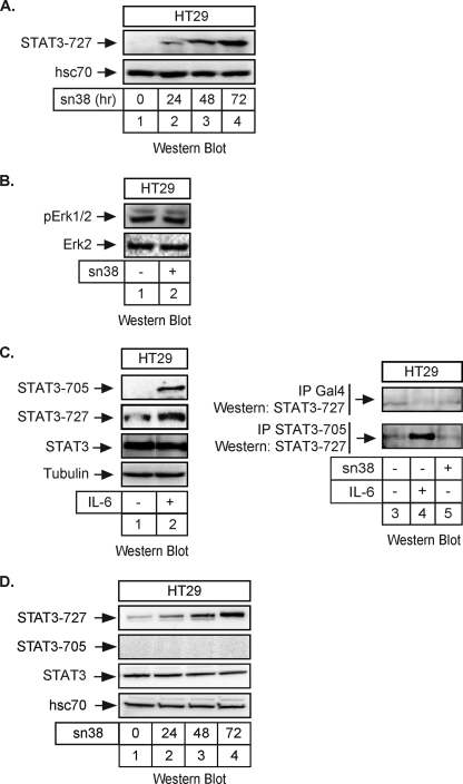
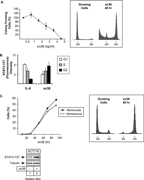
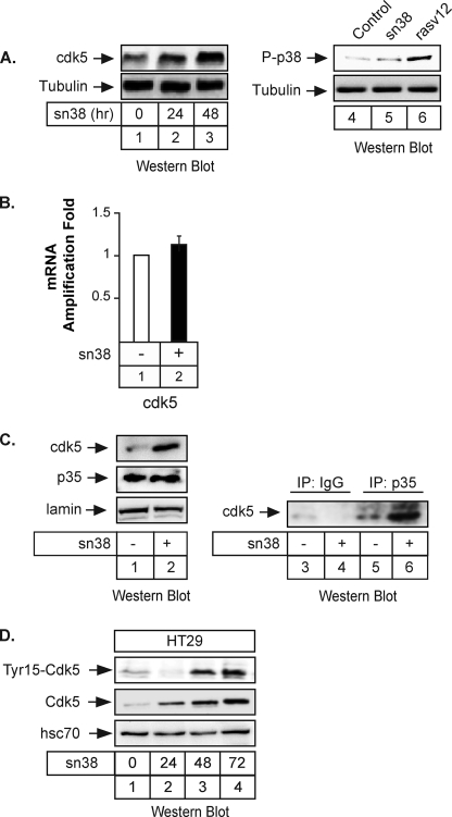
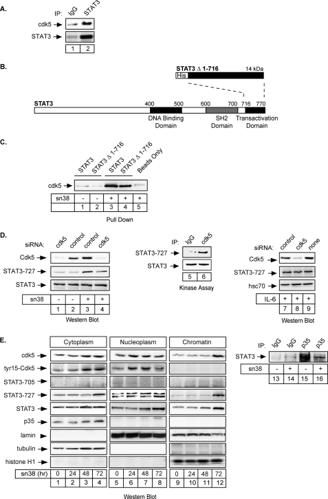
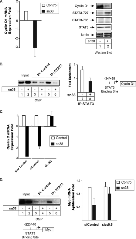
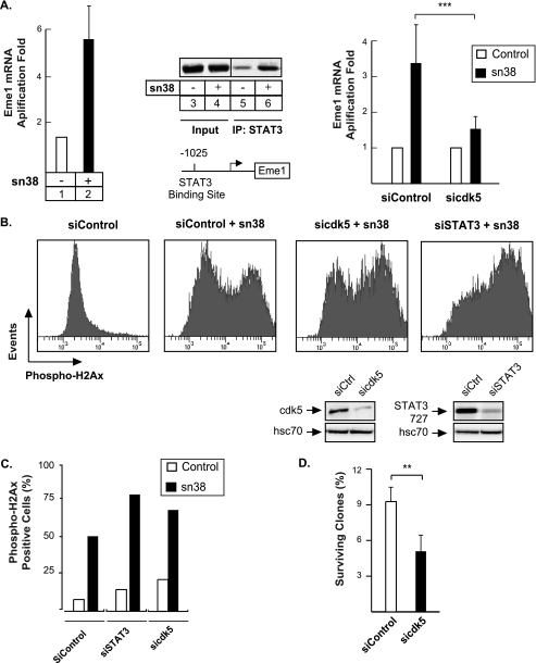
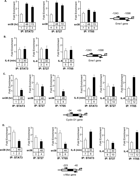
The STAT3 transcription factors are cytoplasmic proteins that induce gene activation in response to growth factor stimulation. Following tyrosine phosphorylation, STAT3 proteins dimerize, translocate to the nucleus, and activate specific target genes involved in cell-cycle progression. Despite its importance in cancer cells, the molecular mechanisms by which this protein is regulated in response to DNA damage remain to be characterized. In this study, we show that STAT3 is activated in response to topoisomerase I inhibition. Following treatment, STAT3 is phosphorylated on its C-terminal serine 727 residue but not on its tyrosine 705 site. We also show that topoisomerase I inhibition induced the up-regulation of the cdk5 kinase, a protein initially described in neuronal stress responses. In co-immunoprecipitations, cdk5 was found to associate with STAT3, and pulldown experiments indicated that it associates with the C-terminal activation domain of STAT3 upon DNA damage. Importantly, the cdk5-STAT3 pathway reduced DNA damage in response to topoisomerase I inhibition through the up-regulation of Eme1, an endonuclease involved in DNA repair. ChIP experiments indicated that STAT3 can be found associated with the Eme1 promoter when phosphorylated only on its serine 727 residue and not on tyrosine 705. We therefore propose that the cdk5-STAT3 oncogenic pathway plays an important role in the expression of DNA repair genes and that these proteins could be used as predictive markers of tumors that will fail to respond to chemotherapy.


Figures

FIGURE 1.
FIGURE 1.
STAT3 is phosphorylated on its serine 727 residue following topoisomerase I inhibition. A, HT29 cells were treated with sn38 (5 ng/ml) or not for the indicated times. Following stimulation, total cell extracts were prepared, and serine 727 phosphorylation was analyzed by Western blot using polyclonal antibodies directed against the phosphorylated form of the protein. The membrane was reprobed with an antibody directed against hsc70 as a loading control (n = 5). Note that, in every experiment, sn38 was added to the cell culture just after cell plating to get an optimal inhibition of cell cycle progression. B, HT29 cells were treated with sn38 (5 ng/ml) for 48 h. Following stimulation, cell extracts were prepared and p-Erk1/2 and Erk2 expression was analyzed by Western blot with polyclonal antibodies directed against these proteins (n = 3). C, HT29 cells were serum-starved for 2 days and stimulated with IL-6 (20 ng/ml) for 30 min. STAT3 activation was analyzed by Western blot using antibodies directed against the different phosphorylated forms of the proteins or against its non-phosphorylated form (n = 2). In parallel, whole cell extracts were immunoprecipitated with polyclonal antibodies directed against the tyrosine-phosphorylated form of STAT3 (−705) or control antibodies (Gal4), and samples were then analyzed by Western blot using polyclonal antibodies directed against STAT3 Ser727 (n = 2). D, HT29 cells were treated with sn38 (5 ng/ml) or not for the indicated times. Following stimulation, total cell extracts were prepared, and the serine 727 and tyrosine 705 phosphorylations were analyzed by Western blot using polyclonal antibodies directed against the phosphorylated forms of the protein. The membrane was reprobed with an antibody directed against STAT3 and then hsc70 as a loading control (n = 3).
FIGURE 2.
FIGURE 2.
STAT3 is phosphorylated on serine 727 during G2 arrest. A, HT29 cells were treated or not with different concentration of sn38 for 10–14 days. Colony formation was then counted using an inverted microscope, and the growth of non-treated cells was set up at 100%. Clonogenic survival was then plotted as a fraction relative to these untreated cells (n = 5 ± S.D.). In parallel, growing HT29 cells were treated with sn38 (5 ng/ml) for 48 h, and DNA content and apoptosis were then evaluated by flow cytometry (n = 5). B, HT29 cells were treated with sn38 (5 ng/ml) or IL-6 (20 ng/ml) as indicated, and DNA content and serine phosphorylation were then analyzed by flow cytometry analysis using polyclonal antibodies directed against the serine 727-phosphorylated form of STAT3 (n = 3). C, growing HCT116 cells were treated or not with sn38 for different times as indicated. The percentage of senescent cells was evaluated as the number of cells expressing SA-β-gal activity and micronuclei (left part, n = 3). In parallel, DNA content was evaluated by flow cytometry after 48 h (right part), and the phosphorylation of STAT3 on its serine residue was analyzed by Western blot as described above (n = 3, bottom).
FIGURE 3.
FIGURE 3.
The cdk5 kinase is activated following topoisomerase I inhibition. A, growing HT29 cells were treated or not with sn38 (5 ng/ml) for 24 or 48 h. Following stimulation, total cell extracts were prepared, and cdk5 expression was analyzed by Western blot using polyclonal antibodies directed against the kinase (lanes 1–3, n = 5). Under the same conditions, the phosphorylation of the p38 kinase was investigated. As a control, cells were transfected with the rasv12 oncogene to induce p38 activation (lanes 4–6, n = 3). The membranes were reprobed with an antibody directed against tubulin as a loading control. B, growing HT29 cells were incubated with sn38 (5 ng/ml) for 48 h, and the expression of the cdk5 mRNA was analyzed by quantitative RT-PCR experiments (n = 3). C, growing HT29 cells were treated as described above, and after 48 h, whole cell extracts were prepared and Western blot analysis was performed with polyclonal antibodies directed against cdk5, p35, or lamin as a loading control (lanes 1 and 2). In parallel, extracts were immunoprecipitated with polyclonal antibodies directed against p35 (lanes 5 and 6) or a control serum (lanes 3 and 4). Samples were then analyzed by Western blot using polyclonal antibodies directed against cdk5 (n = 3). D, HT29 cells were treated with sn38 as described previously, and the expression and phosphorylation of cdk5 on its tyrosine 15 residue were analyzed by Western blot (n = 3).
FIGURE 4.
FIGURE 4.
Cdk5 interacts with STAT3 to induce its phosphorylation on Serine 727. A, HT29 cells were treated with sn38 for 48 h, and whole cell extracts were then immunoprecipitated with polyclonal antibodies directed against STAT3 proteins (lane 2) or a control serum (lane 1), separated by SDS-PAGE, transferred to a nitrocellulose filter, and probed with polyclonal antibodies directed against STAT3 or cdk5 proteins as indicated. B, representation of the fusion proteins used in the pulldown experiments. C, total cell extracts (300 μg) were incubated with histidine, with His-tagged STAT3Cter (STAT3Δ1–716), or with the full-length STAT3 (his-STAT3) immobilized on nickel-agarose beads (400 ng). Samples were then separated on polyacrylamide gels, and cdk5 binding was detected by Western blot using anti-cdk5 polyclonal antibodies (lanes 1–4). D, growing HT29 cells were either transfected with cdk5-specific siRNA oligonucleotides or control oligonucleotides as indicated. Cdk5 expression and phosphorylation of STAT3 on its serine residue were monitored after treatment with sn38 for 48 h (lanes 1–4, n = 4). In parallel, cdk5 was immunoprecipitated from sn38-treated cells and incubated with STAT3Δ1–716 for 10 min at RT in the presence of cold ATP. The phosphorylation of STAT3 on its serine 727 residue was analyzed by Western blot as described above (lanes 5 and 6). In parallel, growing HT29 cells were either transfected with cdk5-specific siRNA or control siRNA as indicated, serum-starved, and then stimulated with IL-6 for 30 min. Cdk5 expression and STAT3 phosphorylation were monitored as above (n = 2). E, HT29 cells were treated with sn38 for the indicated times and cytoplasmic, nuclear, or chromatin extracts were prepared and analyzed by Western blot analysis using antibodies directed against the indicated proteins (n = 2, lanes 1–12). Lamin, tubulin, and histone expression were used as loading controls for each compartment. In parallel, HT29 cells were treated or not with sn38 for 48 h, and whole cell extracts were then immunoprecipitated with polyclonal antibodies directed against p35 (lanes 15 and 16) or a control serum (lanes 13 and 14), separated by SDS-PAGE, transferred to a nitrocellulose filter, and probed with polyclonal antibodies directed against STAT3 proteins as indicated.
FIGURE 5.
FIGURE 5.
Cdk5 is involved in the down-regulation of cyclin D1 and myc following topoisomerase I inhibition. A, growing HT29 cells were incubated with sn38 (5 ng/ml) for 48 h, and the expression of the cyclin D1 mRNA was analyzed by quantitative RT-PCR experiments (n = 3). In parallel, Western blot experiments were also performed to confirm the down-regulation of the cyclin D1 protein and the phosphorylation of STAT3 on its serine residue (lanes 1 and 2). B, HT29 growing cells were treated as described above, and soluble chromatin was prepared from the indicated cells and immunoprecipitated with antibodies directed against STAT3 or control antibodies. DNA was amplified using one pair of primers that covers the STAT3 proximal binding site of the cyclin D1 promoter. ChIP assays were analyzed on agarose gel (left part) or quantified by real-time PCR (n = 3, right part of the figure). C, growing HT29 cells were left untreated or transfected with cdk5-specific or control siRNA oligonucleotides as indicated. Cyclin D1 mRNA expression was analyzed by quantitative RT-PCR experiments following sn38 treatment (n = 3). D, growing HT29 cells were treated as described above, and the association of STAT3 with the myc proximal promoter was analyzed by ChIP (lanes 1–6). In parallel, myc expression was evaluated by quantitative RT-PCR in the presence or absence of cdk5 (right part, n = 4 ± S.D.).
FIGURE 6.
FIGURE 6.
The cdk5-STAT3 pathway regulates the expression of Eme1 and reduces DNA damage. A, Eme1 mRNA expression was analyzed by quantitative RT-PCR (lanes 1 and 2), and STAT3 association with the Eme1 promoter was characterized by ChIP (lanes 3–6) following sn38 treatment. In parallel, cells were transfected with control or cdk5 siRNA and then treated with sn38 (5 ng/ml) for 48 h. The expression of the Eme1 mRNA was analyzed by RT-QPCR experiments (n = 3 ± S.D., p < 0.001). B and C, growing HT29 cells were transfected with specific or control siRNA and treated or not with sn38 (5 ng/ml). The generation of DNA double strand breaks was quantified by FACS analysis using polyclonal antibodies directed against the ser139 phosphorylated form of histone H2Ax (one experiment representative of three). D, HT29 cells were transfected with pools of siRNAs directed against cdk5 or control siRNAs for 48 h. Cells were then split and treated with sn38 for 10–14 days. The percentage of colony-forming cells was evaluated as compared with non-treated cells (n = 3 ± S.D., p < 0.01).
FIGURE 7.
FIGURE 7.
STAT3 is recruited to the Eme1 promoter when phosphorylated only on its serine 727 residue. Growing HT29 cells were treated or not with sn38 as indicated above and soluble chromatin was prepared and immunoprecipitated with antibodies directed against STAT3 (IP:STAT3) or its serine or tyrosine phosphorylated forms (IP:S727 or IP:Y705). In parallel, cells were serum-starved and stimulated or not with IL-6 (10 ng/ml) for 30 min, and the chromatin was immunoprecipitated under the same conditions. DNA was amplified using pair of primers that covers the STAT3 proximal binding sites of the cyclin D1 (panel C), Myc (panel D), and Eme1, (panel A and B) promoters as indicated. ChIP assays were then quantified by real-time PCR as compared with the signals obtained on each genes with a control IgG (n = 3). Note that sn38 (−) in the legend means growing cells, whereas IL6 (0) means serum-starved cells.

References

    1. Bromberg J. (2002) J. Clin. Invest. 109, 1139–1142 - PMC - PubMed
    1. Yu H., Jove R. (2004) Nat. Rev. Cancer 4, 97–105 - PubMed
    1. Levy D. E., Lee C. K. (2002) J. Clin. Invest. 109, 1143–1148 - PMC - PubMed
    1. Bromberg J. F., Horvath C. M., Besser D., Lathem W. W., Darnell J. E., Jr. (1998) Mol. Cell. Biol. 18, 2553–2558 - PMC - PubMed
    1. Bromberg J. F., Wrzeszczynska M. H., Devgan G., Zhao Y., Pestell R. G., Albanese C., Darnell J. E., Jr. (1999) Cell 98, 295–303 - PubMed

Publication types

MeSH terms

LinkOut - more resources