. 2010 Aug 6;285(32):24420-31.
doi: 10.1074/jbc.M110.115444. Epub 2010 Jun 1.

Pannexin1 and Pannexin2 channels show quaternary similarities to connexons and different oligomerization numbers from each other

Affiliations

Pannexin1 and Pannexin2 channels show quaternary similarities to connexons and different oligomerization numbers from each other

Cinzia Ambrosi et al. J Biol Chem. .

Abstract

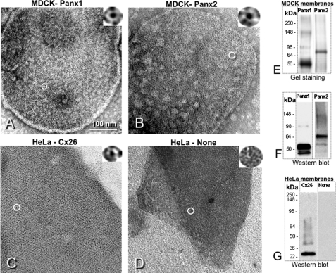
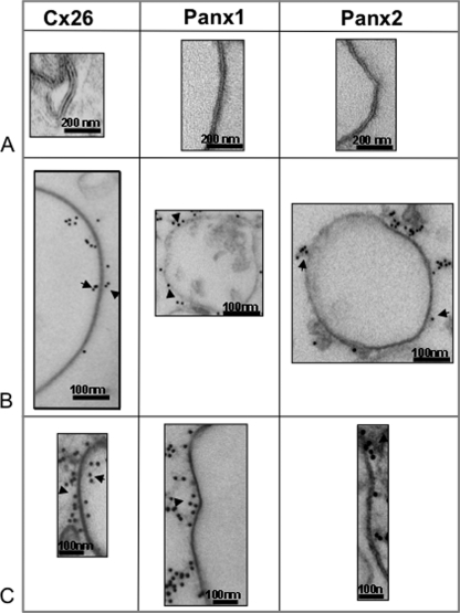
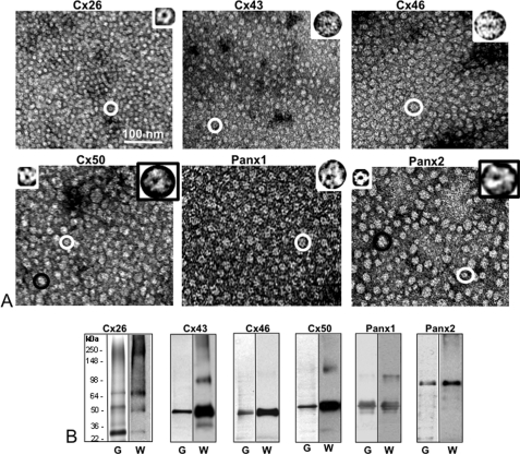
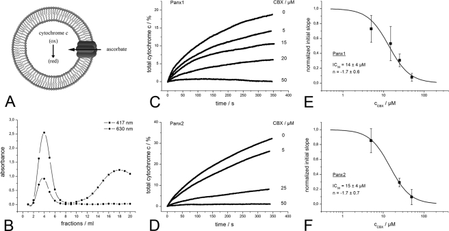
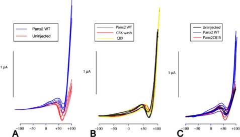
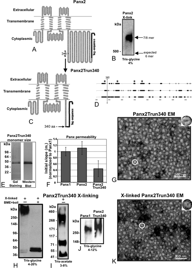
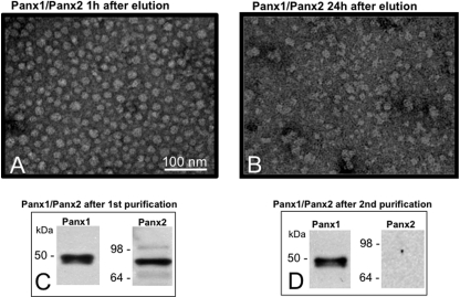
Pannexins are homologous to innexins, the invertebrate gap junction family. However, mammalian pannexin1 does not form canonical gap junctions, instead forming hexameric oligomers in single plasma membranes and intracellularly. Pannexin1 acts as an ATP release channel, whereas less is known about the function of Pannexin2. We purified cellular membranes isolated from MDCK cells stably expressing rat Pannexin1 or Pannexin2 and identified pannexin channels (pannexons) in single membranes by negative stain and immunogold labeling. Protein gel and Western blot analysis confirmed Pannexin1 (Panx1) or Pannexin2 (Panx2) as the channel-forming proteins. We expressed and purified Panx1 and Panx2 using a baculovirus Sf9 expression system and obtained doughnut-like structures similar to those seen previously in purified connexin hemichannels (connexons) and mammalian membranes. Purified pannexons were comparable in size and overall appearance to Connexin46 and Connexin50 connexons. Pannexons and connexons were further analyzed by single-particle averaging for oligomer and pore diameters. The oligomer diameter increased with increasing monomer molecular mass, and we found that the measured oligomeric pore diameter for Panxs was larger than for Connexin26. Panx1 and Panx2 formed active homomeric channels in Xenopus oocytes and in vitro vesicle assays. Cross-linking and native gels of purified homomeric full-length and a C-terminal Panx2 truncation mutant showed a banding pattern more consistent with an octamer. We purified Panx1/Panx2 heteromeric channels and found that they were unstable over time, possibly because Panx1 and Panx2 homomeric pannexons have different monomer sizes and oligomeric symmetry from each other.


Figures

FIGURE 1.
FIGURE 1.
Panx1 and Panx2 show a channel topology in mammalian cell membrane, very similar to gap junction proteins. Membranes isolated from Panx1 (A) and Panx2 (B) exogenously overexpressed in MDCK cells contain channel-like structures similar in appearance to Cx26 exogenously expressed in HeLa cells (C). As a negative control, a membrane isolated from parental HeLa cells shows no channel-like structures (D). Gel staining (E) and Western blot (F) on denaturing PAGE gels show good purity and high specificity of our membrane purifications. A positive control shows a Cx26 band (G) but after stripping and reprobing the same filter with several other antibodies shows no cross-reactivity. The antibodies used in this figure are monoclonal Cx26, our monoclonal Panx1, and polyclonal Panx2 antibodies described under “Experimental Procedures.”
FIGURE 2.
FIGURE 2.
Membrane cross-section comparison between Cx26 and Panx1 or Panx2 showing relevant differences. Membrane profiles of Cx26, Panx1, and Panx2 show double layers for Cx26 and single layer for Panx1 and Panx2 (A). Immunolabeling with specific primary antibodies and with secondary gold conjugated antibodies shows that the gold labels both sides of the membrane profile for Cx26 and only one side for Panx1 and Panx2 (B). An enlarged view of an immunolabeled membrane is shown (C) for easier visualization.
FIGURE 3.
FIGURE 3.
Isolated pannexin oligomers (pannexons) confirm similar features to connexons. Pannexons and connexons expressed and purified from baculovirus infected Sf9 cells. A, EM analysis going from a smaller connexin (Cx26 top left image) to Panx2 (bottom right image). Larger proteins (Panx2 and Cx50) have more heterogeneous morphology of structures, perhaps because of differential staining of different orientations. The images are displayed at the same magnification to show increasing channel size with increasing monomer size. B, stained protein gels (labeled with G for gel on the bottom) and Western blots (labeled with W for Western blot) by the side of each protein demonstrate good purity of these preparations. Note that Western blots can tend to overemphasize dimeric bands. White circles and white boxed insets indicate oligomers that have smaller diameters than ones with black circles and in black box insets.
FIGURE 4.
FIGURE 4.
Panx2 homomeric channels are functional. A, schematic of the cytochrome c-based vesicle assay. If Panx2 channels are functionally reconstituted, ascorbate crosses the lipid bilayer. Intraliposomal cytochrome c gets reduced as monitored at 417 nm. B, fractions of a characteristic size exclusion chromatography to separate free cytochrome c from Panx-1-palmitoyl-2-oleoyl-sn-glycero-3-phosphocholine-proteoliposomes with entrapped cytochrome c. Turbidity of the vesicle suspension is followed at 630-nm, cytochrome c-containing fractions at 417 nm. C, time-resolved reduction of intraliposomal cytochrome c mediated by the transport of ascorbate via a transmembrane spanning active Panx1 channel and inhibition with CBX. D, as in C for Panx2. E, semi-logarithmic plot illustrating the concentration-dependent blocking of the Panx1 pannexons by CBX. Each data point represents the normalized initial slope mi of the Panx1-mediated intraliposomal cytochrome c reduction. The solid line is a fit to the data points using the Hill equation of the form: mi = mi MAX (cCBXn/([IC50]n + cCBXn)) where mi MAX is the initial slope recorded in buffer without CBX (set as unity), cCBX is the drug concentration, IC50 is the concentration giving half of the maximal inhibition, and n is the Hill coefficient. F, as in E for Panx2.
FIGURE 5.
FIGURE 5.
Panx2 channels expressed in Xenopus oocytes opened at high positive membrane potentials and were insensitive to inhibitors. A voltage ramp from −100 mV to +100 mV over a span of 70 s applied to single, uninjected oocytes induced a transient inward current carried by endogenous channels (probably voltage activated sodium channels) (red traces in A). These currents were somewhat variable between oocytes and even within the same oocyte in response to repetitive activation as indicated by the variability of the amplitude and position of peak activity. Expression of Panx2 resulted in large outward currents most prominent at potentials exceeding +75 mV (blue traces). Shown here are five representative traces from each condition. Membrane currents in Panx2-expressing oocytes before (black traces), during (yellow traces), and after washout (red traces) of CBX are shown in B. CBX slightly attenuated the endogenous inward currents and led to larger peak currents as if stimulating rather than inhibiting Panx2 currents, an effect that was not immediately reversed upon washout. This figure contains three traces each for Panx2 WT-injected oocytes and subsequent CBX-treated oocytes and two traces for the CBX wash out oocytes. In C, mutagenesis of cysteines in Panx2 eliminated currents in injected oocytes (see also Table 2). Shown here are five representative traces from the Panx2C81S mutant, Panx2 WT, and uninjected oocytes.
FIGURE 6.
FIGURE 6.
Cross-linking of a Panx2 truncation mutant for stoichiometric analysis reveal nonhexameric assemblies. A, folding diagrams for full-length Panx2. B, cross-linked Panx2 run on a 4% Tris-glycine gel reveals a band located well above a hexamer. This band corresponds to either a heptamer or octamer. To distinguish between an octamer and heptamer stoichiometry, a truncation mutant of Panx2 was constructed so that Panx2 is truncated after Ser340 plus a 30-amino acid tag (370 amino acids total). C, topology diagram for Panx2Trun340-V5-His6. D, predicted secondary structure in the Panx2 C terminus (amino acids 317–674) according to three different prediction algorithms NNPredict (top), GOR4 (middle), and PSIPRED (bottom). Heavy black lines mark stretches of putative α-helices, and heavy gray lines indicate β structure propensity. An asterisk denotes predicted secondary structure elements longer than 3 amino acids common among these three predictions. The dotted arrow indicates the truncation position between Ser340 and Gln341. E, purified Panx2Trun340-V5-His6 preparations were analyzed by gel stainings and Western blots that this protein maps at ∼41 kDa as expected. Truncated Panx2 was cross-linked with DSP 300 μg/ml after purification and analyzed on different gels. F, cytochrome c vesicle permeability measurements comparing Panx1-V5-His6, Panx2 WT V5-His6, and Panx2Trun340 V5-His6 (as in Fig. 4). Comparison of the initial slopes mi of the fitted line to data points recorded during the first 25 s after the addition of ascorbate for the three pannexons indicated that Panx2 truncation pannexons had a reduced permeability to ascorbate. Each category has an n = 3. G, electron micrograph shows channels formed by Panx2Trun340-V5-His6. H, left lane, Panx2 shows the upper band mapping above 250 kDa, suggesting that it is not a hexamer (41 kDa × 6 = 246 kDa). Right lane, cross-linked Panx2 was boiled in the presence of 5% β-mercaptoethanol and shows the monomeric band mapping as expected ∼41 kDa. I, cross-linked Panx2Trun340-V5-His6 is run on a higher resolving Tris-acetate gel system and is separated into the monomer (41 kDa), the dimer (82 kDa), and the upper band mapping between 268 and 460 kDa. J, the cross-linked Panx2Trun340-V5-His6 upper band maps higher than Panx1 hexameric band (∼300 kDa). The measured position of the Panx2Trun340-V5-His6 band is at a position that confirms that the Panx2 oligomer is mostly likely an octamer. K, electron micrograph shows Panx2Trun340-V5-His6 channel appearance after cross-linking. All baculovirus/purified proteins have a C-terminal V5-His6 tag.
FIGURE 7.
FIGURE 7.
Panx1/Panx2 heteromers are highly unstable. A, EM image containing negatively stained Panx1/Panx2 oligomers examined 1 h after purification. B, after 24 h, the channels are barely recognizable. C, the 1-h sample analyzed by Western blot contained both Panx1 and Panx2 bands. D, after buffer exchange and a second nickel-nitrilotriacetic acid affinity column purification, Panx1 bands are evident, but not Panx2.

Similar articles

Cited by

References

    1. D'hondt C., Ponsaerts R., De Smedt H., Bultynck G., Himpens B. (2009) Bioessays 31, 953–974 - PubMed
    1. Panchin Y. V. (2005) J. Exp. Biol. 208, 1415–1419 - PubMed
    1. Penuela S., Bhalla R., Gong X. Q., Cowan K. N., Celetti S. J., Cowan B. J., Bai D., Shao Q., Laird D. W. (2007) J. Cell Sci. 120, 3772–3783 - PubMed
    1. Baranova A., Ivanov D., Petrash N., Pestova A., Skoblov M., Kelmanson I., Shagin D., Nazarenko S., Geraymovych E., Litvin O., Tiunova A., Born T. L., Usman N., Staroverov D., Lukyanov S., Panchin Y. (2004) Genomics 83, 706–716 - PubMed
    1. Jiang H., Zhu A. G., Mamczur M., Falck J. R., Lerea K. M., McGiff J. C. (2007) Br. J. Pharmacol. 151, 1033–1040 - PMC - PubMed

Publication types