. 2010 Aug 6;285(32):24871-81.
doi: 10.1074/jbc.M110.133280. Epub 2010 Jun 1.

Novel polymorphisms of nuclear receptor SHP associated with functional and structural changes

Affiliations

Novel polymorphisms of nuclear receptor SHP associated with functional and structural changes

Taofeng Zhou et al. J Biol Chem. .

Abstract

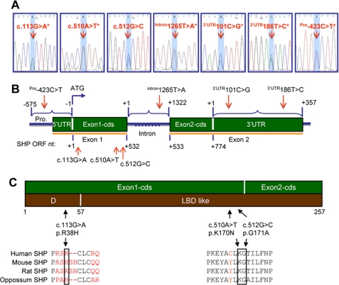
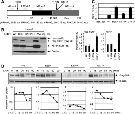
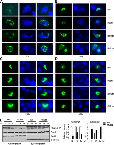
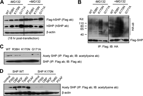
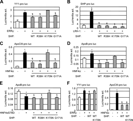
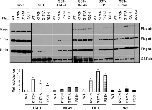
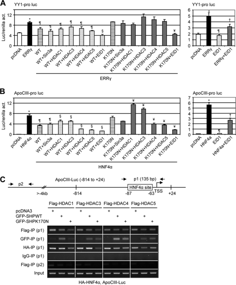
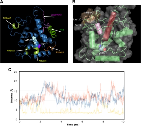
We identified three heterozygous nonsynonymous single nucleotide polymorphisms in the small heterodimer partner (SHP, NROB2) gene in normal subjects and CADASIL (cerebral autosomal dominant arteriopathy with subcortical infarcts and leukoencephalopathy)-like patients, including two novel missense mutations (p.R38H, p.K170N) and one of the previously reported polymorphism (p.G171A). Four novel heterozygous mutations were also identified in the intron ((Intron)1265T-->A), 3'-untranslated region ((3'-UTR)101C-->G, (3'-UTR)186T-->C), and promoter ((Pro)-423C-->T) of the SHP gene. The exonic R38H and K170N mutants exhibited impaired nuclear translocation. K170N made SHP more susceptible to ubiquitination mediated degradation and blocked SHP acetylation, which displayed lost repressive activity on its interacting partners ERRgamma and HNF4alpha but not LRH-1. In contrast, G171A increased SHP mRNA and protein expression and maintained normal function. In general, the interaction of SHP mutants with LRH-1 and EID1 was enhanced. K170N also markedly impaired the recruitment of SHP, HNF4alpha, HDAC1, and HDAC3 to the apoCIII promoter. Molecular dynamics simulations of SHP showed that G171A stabilized the nuclear receptor boxes, whereas K170N promoted the conformational destabilization of all the structural elements of the receptor. This study suggests that genetic variations in SHP are common among human subjects and the Lys-170 residue plays a key role in controlling SHP ubiquitination and acetylation associated with SHP protein stability and repressive function.


Figures

FIGURE 1.
FIGURE 1.
Polymorphisms in the human SHP gene in normal and CADASIL subjects. A, electropherograms show the six novel and one previously identified heterozygous sequence variants. All variants were identified in the forward and reverse genomic DNA strands. The SNP of interest is highlighted in light blue. The asterisks indicate novel variants identified in this study which have not been reported before. B, a diagram of the SHP gene shows the location of the variants. Polymorphisms were identified in exon 1 (c.113G→A*, c.510A→T*, c.512G→C), intron (intron1265T→A*), 3′-UTR (3′-UTR101C→G*, 3′-UTR186T→C*), and promoter (Pro-423C→T) of the SHP gene. Mutation nomenclature was numbered based on GenBankTM cDNA NM_021969.2 and protein NM_068804.1 sequences. In the open reading frame (ORF) of the coding region, nucleotide +1 corresponds to the A of the ATG start codon. In the intron region, nucleotide +1 corresponds to the 1st nucleotide of intron. In the 3′-UTR region, nucleotide +1 corresponds to the 1st nucleotide downstream of the stop codon. In the 5′-UTR and promoter region, nucleotide −1 corresponds to the 1st nucleotide upstream of the start codon. C, a diagram of the SHP gene shows the location of variants that result in substitution of histidine for arginine at position 38 in the SHP hinge region, asparagine for lysine at position 170, and alanine for glycine at position 171 in the SHP ligand-binding domain-like domain.
FIGURE 2.
FIGURE 2.
Expression of mutant SHP proteins and their half-life. A, a diagram shows the structure of SHP and location of R38H, K170N, and G171A. The receptor interaction (Int) and repression (Rep) domains are indicated by black and gray boxes, respectively. The proximal locations of NRbox1, NRbox2, NRbox3, and PSXXLP are indicated. aa, amino acids. B, left, shown is a Western blot analysis of the hSHP protein in Hepa-1 cells that overexpressed with FLAG expression plasmids containing each hSHP mutation (SHPR38H, SHPK170N, and SHPG171A) using anti-FLAG antibodies (ab). The experiments were repeated three times with anti-FLAG antibodies and one time with anti-hSHP antibodies. neg. con., cells transfected with equal amounts of empty vector; no bands with correct size were detected by both antibodies. Right, shown is a quantitative analysis of each band intensity on the left (n = 3; *, p < 0.01 relative to WT). The results were expressed as -fold changes relative to SHPWT, which was set as 1. C, shown is a real-time quantitative PCR analysis of hSHP mRNA in mouse Hepa-1 cells overexpressed with SHPWT, SHPR38H, SHPK170Nv and SHPG171A expression vectors. The results were compared with SHPWT, which was set as 1 (*, p < 0.01). neg. con., cells transfected with equal amounts of pcDNA3 empty vector. D, top, Western blots to determine the half-life of mutant SHP proteins. Hepa-1 cells were transfected with each FLAG-SHP vector then treated with cycloheximide (CHX, 50 μm), and SHP was detected with anti-FLAG antibodies. Bottom, quantitative analysis of each band intensity at the top.
FIGURE 3.
FIGURE 3.
Subcellular localization of SHP mutants in Hepa-1 cells. A–D, GFP-SHPWT, GFP-SHPR38H, GFP-SHPK170N, and GFP-SHPG171A expression vectors were constructed and overexpressed into mouse Hepa-1 cells. The location of GFP-SHP proteins was monitored by green fluorescence under a microscope at 6 (A), 18 (B), 24 (C), and 48 h (D) post-transfection. E, left, Western blots to determine SHP protein in the nuclear and cytosolic extracts in Hepa-1 cells transfected with FLAG-SHPWT and FLAG-SHPK170N expression vectors using anti-FLAG antibodies are shown. β-Actin, poly(ADP-ribose) polymerase (PARP), and α-tubulin were used as markers and internal controls to detect both nuclear and cytosolic (β-actin), nuclear (PARP), or cytosolic proteins (a-tubulin), respectively. Right, a quantitative analysis of each band intensity is shown on the left and expressed as -fold change relative to WT at each time point, which was set as 1. pt, protein.
FIGURE 4.
FIGURE 4.
Ubiquitination (A and B) and acetylation (C and D) assays to determine SHP degradation. A, HeLa cells were transfected with FLAG-SHPWT, FLAG-SHPR38H, FLAG-SHPK170N, or FLAG-SHPG171A expression vectors in the absence (−) or presence of MG132 (+, 5 μm) for 18 h. SHP proteins were determined using anti-FLAG or anti-SHP antibodies (ab) and Western blots. B, HeLa cells were cotransfected with FLAG-SHPWT, FLAG-SHPR38H, FLAG-SHPK170N, or FLAG-SHPG171A expression vectors together with an HA-ubiquitin (HA-ub) plasmid. FLAG-SHP was immunoprecipitated from cell extracts with anti-FLAG antibodies (IP), and ubiquitinated SHP in the immunoprecipitates was detected by Western blots (IB) with anti-HA antibodies. Positions of ubiquitinated SHP proteins are indicated by a dotted line. C, plasmids expressing the indicated SHP mutants were transfected into HeLa cells. Lysates were immunoprecipitated with anti-FLAG antibodies and blotted with anti-acetyllysine antibodies. The blot was then stripped and blotted with anti-FLAG antibodies. Because of the low basal level of SHPK170N, double amounts of FLAG-SHPK170N plasmids were transfected to keep the FLAG-SHPK170N level similar as other SHP proteins. D, plasmids expressing SHPWT and SHPK170N were co-transfected with acetyltransferases p300, CBP, MOF, Tip60, and PCAF into HeLa cells. Acetylation assays was performed as in C.
FIGURE 5.
FIGURE 5.
Functional analysis of SHP mutants. Transcriptional regulation of the three mutations p.R38H, p.K170N, and p.G171A was examined using luciferase reporter assays. act, activity. A, YY1 promoter luciferase reporter was co-expressed with ERRγ and each mutant SHP expression construct in Hepa-1 cells. B, SHP promoter luciferase reporter was co-expressed with LRH-1 and each mutant SHP expression construct in Hepa-1 cells. C, apoCIII promoter luciferase reporter was co-expressed with HNF4a and each mutant SHP expression construct in Hepa-1 cells. D and E, apoB promoter luciferase reporter was co-expressed with HNF4a (D) or a point mutation HNF4aS78D (E) and each mutant SHP expression construct in Hepa-1 cells. F, left, YY1 promoter luciferase reporter was co-expressed with ERRγ in the presence of SHP WT and K170N mutant. Right, apoCIII promoter luciferase reporter was co-expressed with HNF4α in the presence of SHP WT and K170N mutant. Luc activities were determined and normalized to Renilla activities. Data are expressed as the means ± S.D. (*, p < 0.01 versus the white bar; ¶, p < 0.01 versus the black bar; §, p < 0.01 versus the white bar). pro, promoter; luc, luciferase reporter.
FIGURE 6.
FIGURE 6.
GST pulldown assay to determine in vitro interaction of SHP mutants with LRH-1, HNF4a, EID1, and ERRγ. FLAG-SHPWT, FLAG-SHPK170N, FLAG-SHPG171A, and FLAG-SHPR38H were in vitro translated and used to interact with GST fusion proteins GST-LRH-1, GST-HNF4a, GST-EID1, and GST-ERRγ, which were expressed from bacterial E. coli BL21/DE3/RIL. Blots from three different exposure times (5 s, 1 min, and 5 min) are presented. Lower panel, -fold changes relative to WT in each group, which was set as 1 (*, p < 0.01). ab, antibody.
FIGURE 7.
FIGURE 7.
Co-repressor recruitment by SHP mutants. A and B, shown are transient transfection assays to determine the effects of co-repressors on WT and SHPK170N inhibition of YY1 (A) and apoCIII (B) promoter reporters. Luc activities were determined and normalized to Renilla activities (act.). Data are expressed as the means ± S.D. (*, p < 0.01 versus white bar; ¶, p < 0.01 versus black bar; §, p < 0.01 versus WT striped bar; ¥, p < 0.01 versus K170N striped bar; p < 0.01 versus black bar). Pro, promoter; Luc, luciferase reporter. C, chromatin immunoprecipitation assays determine the recruitment of co-repressors on the apoCIII promoter by HNF4a and SHP. HeLa cells were co-transfected with FLAG-HDAC1, -3, -4, and -5, GFP-SHPWT and GFP-SHPK170N, HA-HNF4α, and apoCIII promoter (−814 to +24), and anti-FLAG, anti-GFP, and anti-HA antibodies were used to co-immunoprecipitate each corresponding protein, respectively. Two sets of primers were designed for chromatin immunoprecipitation assays. P1 could detect coimmunoprecipitation (IP) of each protein on exogenously overexpressed apoCIII promoter, whereas p2 was located 4 kb upstream from transcriptional start site (TSS) and, thus, served as a negative control.
FIGURE 8.
FIGURE 8.
Molecular dynamics simulations of SHP mutants. A, locations of the Lys-170 and Gly-171 in the structure of SHP show the structural elements involved in mediating the biological functions of SHP are also shown. B, a salt bridge interaction in the NR2box motif is shown whose formation was promoted by G171A replacement. C, analysis is shown of the Glu-111—Lys-120 salt bridge interaction in SHP wild type (blue line), SHPK170N (red line), and SHPG171A (yellow line) along the 10 ns of molecular dynamic simulation. The salt bridge is considered to be formed if the distance between the carboxylic oxygen atoms of Glu111 and the side chain nitrogen atom of Lys-120 is below a cut-off value of 3.5 Å.

Similar articles

Cited by

References

    1. Markward N. J. (2007) AMIA Annual Symposium Proceedings, Chicago, IL, November 10–14, 2007, p. 1041, American Medical Informatics Association, Bethesda, MD - PubMed
    1. Lee H. K., Lee Y. K., Park S. H., Kim Y. S., Park S. H., Lee J. W., Kwon H. B., Soh J., Moore D. D., Choi H. S. (1998) J. Biol. Chem. 273, 14398–14402 - PubMed
    1. Seol W., Chung M., Moore D. D. (1997) Mol. Cell. Biol. 17, 7126–7131 - PMC - PubMed
    1. Wang L., Liu J., Saha P., Huang J., Chan L., Spiegelman B., Moore D. D. (2005) Cell Metab. 2, 227–238 - PubMed
    1. Huang J., Iqbal J., Saha P. K., Liu J., Chan L., Hussain M. M., Moore D. D., Wang L. (2007) Hepatology 46, 147–157 - PubMed

LinkOut - more resources