

myo-Inositol-1-phosphate synthase is required for polar auxin transport and organ development
- PMID: 20516080
- PMCID: PMC2911297
- DOI: 10.1074/jbc.M110.123661
myo-Inositol-1-phosphate synthase is required for polar auxin transport and organ development
Abstract
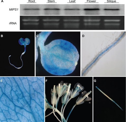
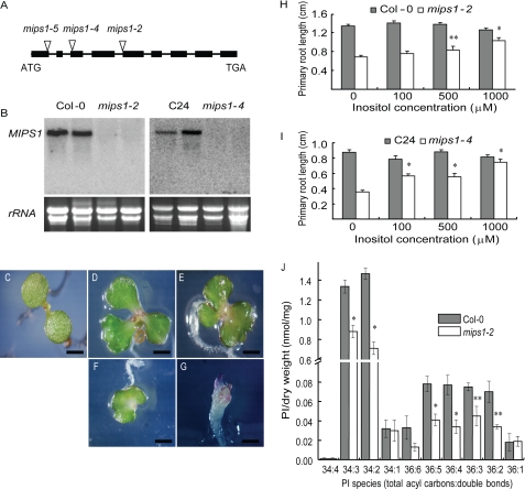
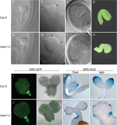
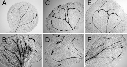
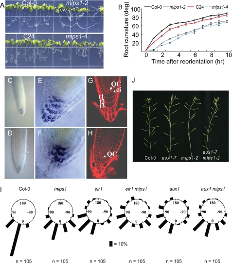
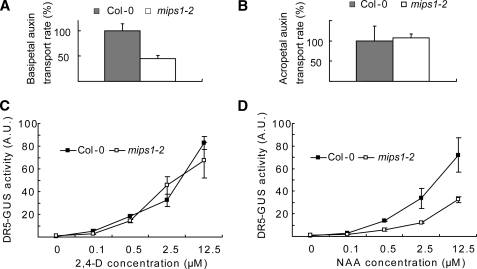
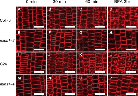
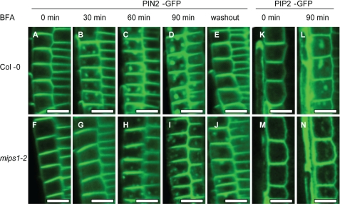
myo-Inositol-1-phosphate synthase is a conserved enzyme that catalyzes the first committed and rate-limiting step in inositol biosynthesis. Despite its wide occurrence in all eukaryotes, the role of myo-inositol-1-phosphate synthase and de novo inositol biosynthesis in cell signaling and organism development has been unclear. In this study, we isolated loss-of-function mutants in the Arabidopsis MIPS1 gene from different ecotypes. It was found that all null mips1 mutants are defective in embryogenesis, cotyledon venation patterning, root growth, and root cap development. The mutant roots are also agravitropic and have reduced basipetal auxin transport. mips1 mutants have significantly reduced levels of major phosphatidylinositols and exhibit much slower rates of endocytosis. Treatment with brefeldin A induces slower PIN2 protein aggregation in mips1, indicating altered PIN2 trafficking. Our results demonstrate that MIPS1 is critical for maintaining phosphatidylinositol levels and affects pattern formation in plants likely through regulation of auxin distribution.
Figures
Similar articles
-
D-myo-inositol-3-phosphate affects phosphatidylinositol-mediated endomembrane function in Arabidopsis and is essential for auxin-regulated embryogenesis.Plant Cell. 2011 Apr;23(4):1352-72. doi: 10.1105/tpc.111.083337. Epub 2011 Apr 19. Plant Cell. 2011. PMID: 21505066 Free PMC article.
-
The Arabidopsis thaliana Myo-inositol 1-phosphate synthase1 gene is required for Myo-inositol synthesis and suppression of cell death.Plant Cell. 2010 Mar;22(3):888-903. doi: 10.1105/tpc.109.071779. Epub 2010 Mar 9. Plant Cell. 2010. PMID: 20215587 Free PMC article.
-
A Cotton (Gossypium hirsutum) Myo-Inositol-1-Phosphate Synthase (GhMIPS1D) Gene Promotes Root Cell Elongation in Arabidopsis.Int J Mol Sci. 2019 Mar 11;20(5):1224. doi: 10.3390/ijms20051224. Int J Mol Sci. 2019. PMID: 30862084 Free PMC article.
-
Complex regulation of Arabidopsis AGR1/PIN2-mediated root gravitropic response and basipetal auxin transport by cantharidin-sensitive protein phosphatases.Plant J. 2005 Apr;42(2):188-200. doi: 10.1111/j.1365-313X.2005.02369.x. Plant J. 2005. PMID: 15807782
-
1L-myo-inositol-1-phosphate synthase.Biochim Biophys Acta. 1997 Sep 4;1348(1-2):245-56. doi: 10.1016/s0005-2760(97)00122-7. Biochim Biophys Acta. 1997. PMID: 9370339 Review.
Cited by
-
Probing myo-inositol 1-phosphate synthase with multisubstrate adducts.Org Biomol Chem. 2012 Dec 28;10(48):9601-19. doi: 10.1039/c2ob26577j. Epub 2012 Nov 7. Org Biomol Chem. 2012. PMID: 23132282 Free PMC article.
-
Inositol hexakisphosphate biosynthesis underpins PAMP-triggered immunity to Pseudomonas syringae pv. tomato in Arabidopsis thaliana but is dispensable for establishment of systemic acquired resistance.Mol Plant Pathol. 2020 Mar;21(3):376-387. doi: 10.1111/mpp.12902. Epub 2019 Dec 26. Mol Plant Pathol. 2020. PMID: 31876373 Free PMC article.
-
Phosphatidylinositides regulate the cell plate morphology transition during cytokinesis in Arabidopsis.Nat Commun. 2025 Jul 30;16(1):7007. doi: 10.1038/s41467-025-62067-4. Nat Commun. 2025. PMID: 40738881 Free PMC article.
-
Jasmonates-Mediated Rewiring of Central Metabolism Regulates Adaptive Responses.Plant Cell Physiol. 2019 Dec 1;60(12):2613-2620. doi: 10.1093/pcp/pcz181. Plant Cell Physiol. 2019. PMID: 31529102 Free PMC article.
-
Alteration in expression of hormone-related genes in wild emmer wheat roots associated with drought adaptation mechanisms.Funct Integr Genomics. 2011 Dec;11(4):565-83. doi: 10.1007/s10142-011-0231-6. Epub 2011 Jun 8. Funct Integr Genomics. 2011. PMID: 21656015
References
-
- Michell R. H. (2008) Nat. Rev. Mol. Cell Biol. 9, 151–161 - PubMed
-
- Martin T. F. (1998) Annu. Rev. Cell Dev. Biol. 14, 231–264 - PubMed
-
- Roth M. G. (2004) Physiol. Rev. 84, 699–730 - PubMed
-
- Stevenson J. M., Perera I. Y., Heilmann I., Persson S., Boss W. F. (2000) Trends Plant Sci. 5, 252–258 - PubMed
-
- Tan X., Calderon-Villalobos L. I., Sharon M., Zheng C., Robinson C. V., Estelle M., Zheng N. (2007) Nature 446, 640–645 - PubMed
Publication types
MeSH terms
Substances
LinkOut - more resources
Full Text Sources
Molecular Biology Databases
Miscellaneous
