. 2010 Aug 13;285(33):25554-69.
doi: 10.1074/jbc.M110.136622. Epub 2010 Jun 1.

Affinity purification of the Arabidopsis 26 S proteasome reveals a diverse array of plant proteolytic complexes

Affiliations

Affinity purification of the Arabidopsis 26 S proteasome reveals a diverse array of plant proteolytic complexes

Adam J Book et al. J Biol Chem. .

Abstract

Selective proteolysis in plants is largely mediated by the ubiquitin (Ub)/proteasome system in which substrates, marked by the covalent attachment of Ub, are degraded by the 26 S proteasome. The 26 S proteasome is composed of two subparticles, the 20 S core protease (CP) that compartmentalizes the protease active sites and the 19 S regulatory particle that recognizes and translocates appropriate substrates into the CP lumen for breakdown. Here, we describe an affinity method to rapidly purify epitope-tagged 26 S proteasomes intact from Arabidopsis thaliana. In-depth mass spectrometric analyses of preparations generated from young seedlings confirmed that the 2.5-MDa CP-regulatory particle complex is actually a heterogeneous set of particles assembled with paralogous pairs for most subunits. A number of these subunits are modified post-translationally by proteolytic processing, acetylation, and/or ubiquitylation. Several proteasome-associated proteins were also identified that likely assist in complex assembly and regulation. In addition, we detected a particle consisting of the CP capped by the single subunit PA200 activator that may be involved in Ub-independent protein breakdown. Taken together, it appears that a diverse and highly dynamic population of proteasomes is assembled in plants, which may expand the target specificity and functions of intracellular proteolysis.


Figures

FIGURE 1.
FIGURE 1.
Arabidopsis PAG1 gene structure and mutant rescue. A, diagram of the single PAG1 gene encoding the α7 subunit of the CP and location of the pag1-1 T-DNA insertion. Boxes indicate exons, and lines indicate introns. aa, amino acids. B, expression of the PAG1-FLAG protein in homozygous pag1-1 mutants. Crude extracts from WT plants and heterozygous (±) and homozygous (−/−) pag1-1 plants expressing the PAG1-FLAG transgene were subjected to SDS-PAGE and immunoblot analysis with antibodies against PAG1, FLAG, and RPN5a (loading control). C, rescue of homozygous pag1-1 plants with the PAG1-FLAG transgene. Shown are 6-week-old plants homozygous for both the pag1-1 mutation and the PAG1-FLAG transgene grown under a long day photoperiod (16 h light/8 h dark). Bar represents 12 mm. D and E, assembly of the PAG1-FLAG protein into proteasome complexes. Crude extracts from WT and homozygous PAG1-FLAG pag1-1 plants were fractionated by glycerol gradient centrifugation and then assayed enzymatically for CP peptidase activity (D) and for various proteasome subunits by immunoblot analysis (E). Sedimentation positions of the 26 S proteasome, free RP, and the PA200-CP (PA-RP) complexes are indicated by the brackets. F, efficacy of protease inhibitors in stabilizing the PAG1-FLAG protein in vitro. Crude extracts from 10-day-old homozygous PAG1-FLAG pag1-1 seedlings were incubated for 2 h at 25 °C with various protease inhibitors and then analyzed by immunoblot analysis with anti-PAG1 antibodies. The positions of the PAG1-FLAG and PAG1 minus the FLAG tag are indicated. PMSF, phenylmethylsulfonyl fluoride.
FIGURE 2.
FIGURE 2.
Affinity purification of proteasomes from PAG1-FLAG pag1-1 plants. A, SDS-PAGE analysis of the affinity purification steps. Total protein extracted from 10-day-old WT and PAG1-FLAG pag1-1 plants was incubated with anti-FLAG affinity resin, washed, and competitively eluted with the FLAG peptide. The procedure was performed in the presence or absence of ATP. The input, unbound, washed, and eluted fractions were subjected to SDS-PAGE, and the gel was stained for protein with silver. The arrow and closed arrowhead locate the PA200 and the PAG1-FLAG proteins, respectively. The open arrowhead identifies nitrilase, which is nonspecifically enriched during the purification. B, salt dissociation of the 26 S proteasome into the RP and CP subcomplexes. 26 S proteasomes were bound to the anti-FLAG resin in the presence of ATP and either eluted with the FLAG peptide or first eluted with 800 mm NaCl, followed by elution with the FLAG peptide. The brackets locate subunits of the CP and RP subcomplexes. The arrow indicates PA200. C, immunoblot detection of various proteasome subunits in the affinity-purified preparations shown in A. Subunits tested include the CP subunits PAG1 and PBA1, and the RP subunits RPT2, RPN1, RPN5, RPN10, and RPN12a. Other proteins tested include PA200, the CSN4 and CSN5 subunits of the CSN complex, the eIF3-e subunit of the eIF3 complex, HSC70, Rubisco small subunit, and nitrilase (NIT1). D, peptidase activity of affinity-purified proteasomes. Peptidase activity in the presence or absence of MG132 was measured in the crude extract (Cr), and in preparations purified in the presence of ATP, using the substrate succinyl-Leu-Leu-Val-Tyr-7-amido-4-methylcoumarin. Activities were normalized to total protein concentration.
FIGURE 3.
FIGURE 3.
Separation of the various proteasome complexes by native PAGE. A, proteasomes affinity-purified from PAG1-FLAG pag1-1 plants were fractioned by native PAGE in the presence of ATP. Gels were either stained for total protein with silver (Protein) or subjected to immunoblot analysis with antibodies against RPN5, RPN10, PBA1, and PA200 (PA). Migration of the CP, the PA-CP complex, the RP, and singly and doubly capped 26 S proteasomes (26 S-1C and 26 S-2C) are indicated. The ? identifies a larger complex that could represent the core 26 S proteasome associated with additional factors. B, proteasomes separated as in A and then subjected to denaturing SDS-PAGE in the second dimension. A representative native PAGE separation is provided above to help orient the complexes. PA200 and the collection of subunits from the RP and CP subcomplexes are located.
FIGURE 4.
FIGURE 4.
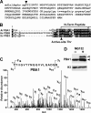
N-terminal processing of the CP β1 subunit PBA1. A, amino acid sequence coverage by MS/MS of PBA1 present in proteasomes affinity-purified from PAG1-FLAG pag1-1 plants. The uppercase residues signify those assigned to specific tryptic peptides (85% coverage for the initial translation product). Lowercase italic residues signify regions not identified in the peptide sequences. Arrowhead locates the most N-terminal residue identified. B, amino acid sequence alignment of the N-terminal region of β1 subunits from A. thaliana (At PBA1), H. sapiens (Hs PSB5), and S. cerevisiae (Sc Pre3). Identical and similar amino acids are show in the black and gray boxes, respectively. Dots denote gaps. The arrowhead marks the N-terminal Thr active site. The bracket locates the most N-terminal peptide identified by MS from trypsinized At PBA1. C, MS/MS spectrum of the N-terminal peptide of PBA1 (residues 13–31) identified by the bracket in B. Parent peptide MH+ = 1894.1 with an Xcorr score of 4.59. The locations of signature b and y ions are indicated along with the calculated peptide sequence. D, unprocessed PBA1 accumulates in Arabidopsis seedlings treated with MG132. Crude extracts were prepared from wild-type plants with or without a 30-h exposure to 100 μm MG132 and then immunoblotted with anti-PBA1 or anti-histone 3 (H3) antibodies (loading control). The open and closed arrowheads identify the unprocessed and mature forms of PBA1, respectively.
FIGURE 5.
FIGURE 5.
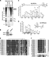
RPN1a and RPN2 assembled into the 26 S proteasome are ubiquitylated. A, extracts from 10-day-old WT and PAG1-FLAG pag1-1 plants were subjected to the affinity purification method for proteasomes in the presence or absence of ATP (see Fig. 2A). The input and eluted fractions were subjected to SDS-PAGE and immunoblot analysis with anti-Ub antibodies. Arrowheads identify poly-Ub trimers and tetramers and the approximate migration position of RPN1 and RPN2. B, co-migration of RPN1 with a ubiquitylated species in affinity-purified proteasomes. Adjacent gel lanes were subjected to immunoblot analysis with anti-Ub or anti-RPN1 antibodies. The arrowheads identify RPN1 and the approximate migration positions of RPN2. C, ESI-MS/MS spectrum of a tryptic peptide from RPN1a (MH+ = 3636.78 m/z) that contains a Gly-Gly footprint derived from Ub attached to Lys-218. D, ESI-MS/MS spectrum of a tryptic peptide from RPN2a/b (MH+ = 1327.80 m/z) that contains a Gly-Gly footprint derived from Ub attached to Lys-165. E and F, amino acid sequence comparison of RPN1 (E) and RPN2 (F) from various eukaryotes at the region surrounding the ubiquitylation sites identified in the Arabidopsis proteins. Identical and similar residues are shown in black and gray boxes, respectively. Dots denote gaps. A. thaliana, At; A. lyrata, Al; G. max, Gm; P. trichocarpa, Pt; O. sativa, Os; Sorghum bicolor, Sb; Brachypodium distachyon Bd; S. moellendorffii, Sm; C. reinhardtii, Cr; P. patens, Pp; Caenorhabditis elegans, Ce; D. melanogaster, Dm; and S. cerevisiae, Sc. The bracket locates the ubiquitylated peptide detected by ESI-MS/MS with the asterisk marking the Lys containing the Ub footprint (Gly-Gly). The vertical line identifies sequences containing the conserved ubiquitylated Lys.
FIGURE 6.
FIGURE 6.
Molecular and genetic analyses of Arabidopsis PA200. A, diagram of the Arabidopsis PA200 gene. Yellow and white boxes denote coding and 3′-untranslated region exons, respectively. Lines show introns. Coding regions for the HEAT repeats and nuclear localization signal (NLS) are in blue and green, respectively. T-DNA insertion sites for the pa200-1–6 mutants are shown in red. Arrows locate the positions of the primers used for RT-PCR analysis in B. aa, amino acids. B, RT-PCR analysis of 2-week-old pa200-2 and pa200-3 seedlings. Total RNA isolated from WT and mutant seedlings was subjected to RT-PCR using the PA200 primers located by the arrows in A. A primer pair specific to PAE2 was used as an internal control. C, immunoblot analyses of total protein from 1-week-old WT and pa200-2 and pa200-3 mutant seedlings with anti-PA200 and anti-UBC1 (loading control) antibodies. D, 10-day-old etiolated pa200-2 and pa200-3 seedling as compared with WT and rpn12a-1 seedlings. E, 12-day-old green WT, pa200-2, pa200-3, and rpn12a-1 seedlings grown under a long-day photoperiod (16 h light/8 h dark). F, immunoblot analysis of Ub-conjugate levels. Seedlings were either treated with DMSO or 100 μm MG132 dissolved in DMSO. Equivalent amounts of total protein were subjected to SDS-PAGE and immunoblotted with anti-Ub antibodies and confirmed by probing with anti-histone 3 (H3) antibodies. Arrowheads indicated free Ub, Ub dimers, trimers, and tetramers. The bracket indicates Ub conjugates. G, increased PA200 protein levels in response to proteasome inhibition. Four-day-old WT, pa200-2, and rpn12a-1 seedlings were treated for 30 h with DMSO or 100 μm MG132 dissolved in DMSO. Equivalent amounts of total protein were subjected to SDS-PAGE and immunoblotted with antibodies against PA200, RPN5a, RPN12a, RPT2a, and PBA1, and HSP70 (loading control). The open and closed arrowheads identify the unprocessed and mature forms of PBA1, respectively. H, PAG1-FLAG affinity purification of proteasomes from 10-day-old seedlings treated with or without 100 μm MG132 for 30 h. The arrowhead locates PA200, the identity of which was confirmed by MS/MS analysis of the excised gel slice.
FIGURE 7.
FIGURE 7.
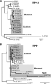
Phylogenetic analysis of the proteasome subunits RPN2 (A) and RPT1 (B). Bayesian phylogenetic analyses were performed using protein sequences from A. thaliana (At), A. lyrata (Al), G. max (Gm), P. trichocarpa (Pt), O. sativa (Os), S. bicolor (Sb), B. distachyon (Bd), S. moellendorffii (Sm), C. reinhardtii (Cr), P. patens (Pp), C. elegans (Ce), D. melanogaster (Dm), H. sapiens (Hs), and S. cerevisiae (Sc). Eudicot- and monocot-specific subclades are highlighted by the shaded boxes. The unique At/Al subclade for RPT1b is also highlighted. Posterior probabilities were calculated from 1000 generations, and values are indicated at each branch node. Trees were rooted with obvious non-plant orthologs. The scale bar represents the average number of substitutions per site.

References

    1. Vierstra R. D. (2009) Nat. Rev. Mol. Cell Biol. 10, 385–397 - PubMed
    1. Finley D. (2009) Annu. Rev. Biochem. 78, 477–513 - PMC - PubMed
    1. Smalle J., Vierstra R. D. (2004) Annu. Rev. Plant Biol. 55, 555–590 - PubMed
    1. Fu H., Reis N., Lee Y., Glickman M. H., Vierstra R. D. (2001) EMBO J. 20, 7096–7107 - PMC - PubMed
    1. Rabl J., Smith D. M., Yu Y., Chang S. C., Goldberg A. L., Cheng Y. (2008) Mol. Cell 30, 360–368 - PMC - PubMed

Publication types

MeSH terms

Substances