. 2010 Jul 1;70(13):5618-27.
doi: 10.1158/0008-5472.CAN-09-3740. Epub 2010 Jun 1.

Atoh1 inhibits neuronal differentiation and collaborates with Gli1 to generate medulloblastoma-initiating cells

Affiliations

Atoh1 inhibits neuronal differentiation and collaborates with Gli1 to generate medulloblastoma-initiating cells

Olivier Ayrault et al. Cancer Res. .

Abstract

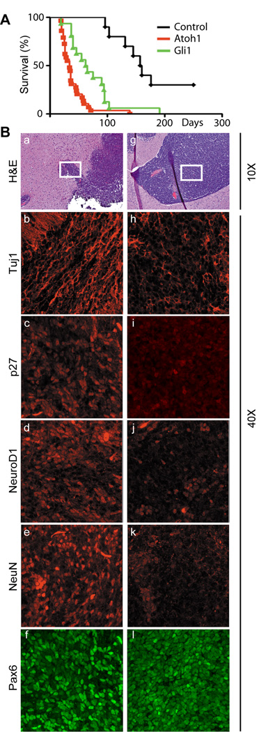
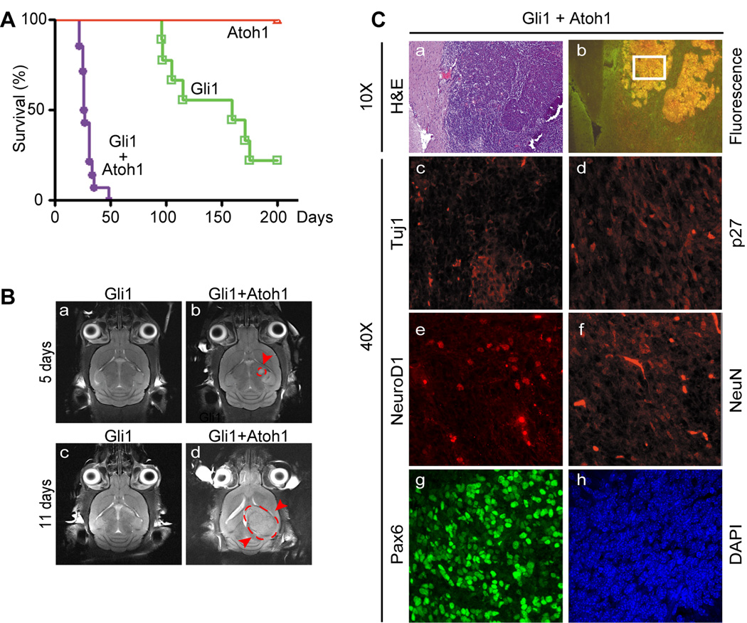
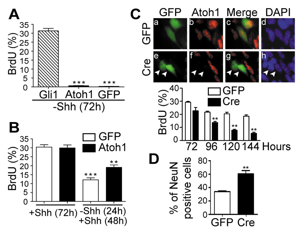
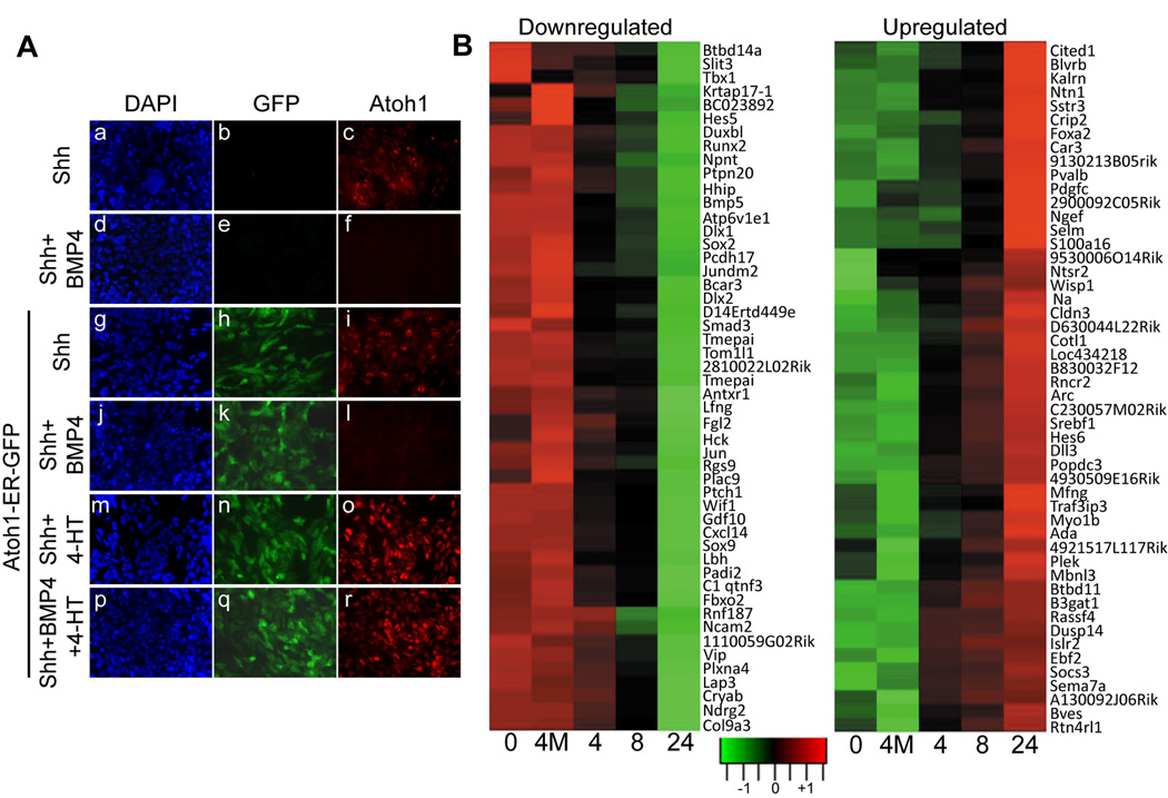
The morphogen and mitogen Sonic Hedgehog (Shh) activates a Gli1-dependent transcription program that drives proliferation of granule neuron progenitors (GNP) within the external germinal layer of the postnatally developing cerebellum. Medulloblastomas with mutations activating the Shh signaling pathway preferentially arise within the external germinal layer, and the tumor cells closely resemble GNPs. Atoh1/Math1, a basic helix-loop-helix transcription factor essential for GNP histogenesis, does not induce medulloblastomas when expressed in primary mouse GNPs that are explanted from the early postnatal cerebellum and transplanted back into the brains of naïve mice. However, enforced expression of Atoh1 in primary GNPs enhances the oncogenicity of cells overexpressing Gli1 by almost three orders of magnitude. Unlike Gli1, Atoh1 cannot support GNP proliferation in the absence of Shh signaling and does not govern expression of canonical cell cycle genes. Instead, Atoh1 maintains GNPs in a Shh-responsive state by regulating genes that trigger neuronal differentiation, including many expressed in response to bone morphogenic protein-4. Therefore, by targeting multiple genes regulating the differentiation state of GNPs, Atoh1 collaborates with the pro-proliferative Gli1-dependent transcriptional program to influence medulloblastoma development.


Figures

Figure 1
Figure 1
Atoh1 accelerates MB formation and inhibits neuronal differentiation. (A) Survival curves of orthotopically transplanted mice that received GNPs from tumor-prone Ptch1+/−;Cdkn2c−/− animals (black) or those infected with vectors encoding Atoh1/RFP (red) or Gli1/GFP (green). (B) MBs accelerated by Gli1 (panels a–f) or Atoh1 (panels g–l) were visualized by H&E staining (panels a and g), by immunofluorescence for the neuronal markers indicated at the left of panels b–f and h–l,. Magnifications are indicated on the right side of the panels.
Figure 2
Figure 2
Atoh1 collaborates with Gli to accelerate MB formation and inhibits neuronal differentiation. (A) Survival curves of orthotopically transplanted mice that received primary GNPs explanted from healthy donors. Cells were engineered to express Atoh1/RFP (red), Gli1/GFP (green) or both (purple). (B) Magnetic resonance images indicating incipient MBs (red arrows and broken line surrounding the tumor) triggered by GNPs co-expressing Atoh1 and Gli1. Donor cells used in this experiment are indicated by asterisks in Table 1. (C) MBs visualized by H&E staining (a) were dually fluorescent (b) and expressed relatively low levels of neuronal markers of differentiation (c–f). Tumor cells were marked by Pax6 (g) and DAPI (h) to visualize nuclei. Magnifications of micrographs are indicated on the left side of the panels.
Figure 3
Figure 3
Atoh1 maintains GNPs in the division cycle but does not directly stimulate proliferation. (A) GNPs expressing Gli1/GFP, Atoh1/GFP, or GFP alone were deprived of Shh for 72 hrs, pulsed for 1.5 hrs with BrdU, and the percentage of GFP+ cells that incorporated BrdU was determined by immunofluorescence. (B) GNPs expressing GFP alone (white bars) or Atoh1/GFP (black bars) were either maintained in Shh for 72 hrs and labeled with BrdU as above or were deprived of Shh for 24 hrs, restimulated for 48 hrs, and then labeled. (C, top) P7 GNPs from “floxed” Atoh1 mice engineered to express GFP alone (panels a–d) or Cre/GFP (panels e–h) (indicated at the left) and cultured in the continued presence of Shh were scored for GFP fluorescence (panels a and e), expression of the endogenous Atoh1 protein by immunofluorescence (panels b and f) (red), or stained with DAPI (panels d and h) (blue). Panels c and g show merged images of panels a and b and of panels e and f, respectively. Cells expressing Cre noted by arrowheads no longer expressed Atoh1. (C, bottom) GNPs infected with GFP control (white bars) or Cre/GFP (black bars) vectors were cultured in presence of Shh for the indicated times (abscissa) and pulsed with BrdU for 1.5 hrs. (D) GNPs studied in panel C were immunostained for NeuN 120 hrs after infection. For all panels, (***) indicates P < 0.001 and (**) P < 0.01 (Student’s t-test).
Figure 4
Figure 4
Downregulation of endogenous Atoh1 protein by BMP4 is overridden by induced Atoh1-ER. (A) Untransduced P7 GNPs (panels a–f) or GNPs expressing Atoh1-ER/GFP (panels g–r) were cultured for 24 hrs in Shh with or without BMP4 and/or 4-HT, as indicated at the left of the panels. Cell nuclei were stained with DAPI (blue). Vector-encoded GFP was visualized by direct fluorescence (green) and Atoh1 by indirect immunofluorescence (red). (B) GNPs transduced with Atoh1-ER were cultured for an additional 24 hours in Shh and BMP4 to downregulate endogenous Atoh1 expression. 4-HT was added for various times in hours (abscissa) before the conclusion of the experiment to induce Atoh1-ER. As a control, some GNPs were infected with a vector encoding an Atoh1 DNA binding mutant that is transcriptionally inactive (4M). RNAs harvested simultaneously from the cultured cells were used to perform microarray gene expression analysis, illustrated by the “heat maps”. Data obtained for the top 50 downregulated and top 50 up-regulated genes are illustrated. Responsive genes showing greater than two-fold changes in expression from a total of 189 unique genes that were assigned to functional categories (P < 0.001) are listed in Supplementary Table 2.

Similar articles

Cited by

References

    1. Sillitoe RV, Joyner AL. Morphology, molecular codes, and circuitry produce the three-dimensional complexity of the cerebellum. Annu Rev Cell Dev Biol. 2007;23:549–577. - PubMed
    1. Wechsler-Reya R, Scott MP. The developmental biology of brain tumors. Annu Rev Neurosci. 2001;24:385–428. - PubMed
    1. Lee Y, Miller HL, Jensen P, et al. A molecular fingerprint for medulloblastoma. Cancer Res. 2003;63:5428–5437. - PubMed
    1. Oliver TG, Read TA, Kessler JD, et al. Loss of patched and disruption of granule cell development in a pre-neoplastic stage of medulloblastoma. Development. 2005;132:2425–2439. - PubMed
    1. Eberhart CG. Even cancers want commitment: lineage identity and medulloblastoma formation. Cancer Cell. 2008;14:105–107. - PMC - PubMed

Publication types

MeSH terms

Substances