Review
. 2010 Jun;2(6):a000919.
doi: 10.1101/cshperspect.a000919. Epub 2010 Feb 10.

The tumor suppressor p53: from structures to drug discovery

Affiliations
Review

The tumor suppressor p53: from structures to drug discovery

Andreas C Joerger et al. Cold Spring Harb Perspect Biol. 2010 Jun.

Abstract

Even 30 years after its discovery, the tumor suppressor protein p53 is still somewhat of an enigma. p53's intimate and multifaceted role in the cell cycle is mirrored in its equally complex structural biology that is being unraveled only slowly. Here, we discuss key structural aspects of p53 function and its inactivation by oncogenic mutations. Concerted action of folded and intrinsically disordered domains of the highly dynamic p53 protein provides binding promiscuity and specificity, allowing p53 to process a myriad of cellular signals to maintain the integrity of the human genome. Importantly, progress in elucidating the structural biology of p53 and its partner proteins has opened various avenues for structure-guided rescue of p53 function in tumors. These emerging anticancer strategies include targeting mutant-specific lesions on the surface of destabilized cancer mutants with small molecules and selective inhibition of p53's degradative pathways.
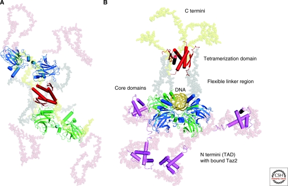

Figures

Figure 1.
Figure 1.
Domain structure of p53. p53 contains a natively unfolded amino-terminal transactivation domain (TAD), which can be further subdivided into the subdomains TAD1 and TAD2, followed by a proline-rich region (PRR). The structured DNA-binding and tetramerization domains (OD) are connected through a flexible linker region. Similarly to the TAD region, the regulatory domain at the extreme carboxyl terminus (CTD) is also intrinsically disordered. The vertical bars indicate the relative missense-mutation frequency in human cancer for each residue based on the TP53 Mutation Database of the International Agency for Research on Cancer (www-p53.iarc.fr) (Petitjean et al. 2007), showing that most cancer mutations are located in the DNA-binding domain. The structure of the DNA-binding domain (PDB code 1TSR) (Cho et al. 1994) is shown as a ribbon representation and colored with a rainbow gradient from the amino terminus (blue) to the carboxyl terminus (red). Sites of cancer hotspot mutations and essential DNA contacts are shown as stick models. Parts of the figure were adapted from Joerger and Fersht (2008).
Figure 2.
Figure 2.
Sequence-specific DNA binding of p53. (A) Cartoon representation of a core domain tetramer bound to DNA, as observed in the crystal structure of human p53 core domain in complex with palindromic half-site DNA (PDB code 2AHI) (Kitayner et al. 2006). Two different views are shown, with individual core domains depicted in different colors: view onto the core domain tetramer, showing core domain-core domain contacts (left) and view along the DNA helix axis (right). (B) Stereo view of the major-groove interaction network of the structure shown in panel A (PDB code 2AHI, chain D). DNA-contact residues are shown as green stick models, and a crucial structural water molecule is shown as a magenta sphere. The polar interaction network involving DNA-contact residues is highlighted with black dashed lines. The orange dashed line indicates hydrophobic interaction between the Cβ atom of Ala276 and a thymine base. Nucleotides are numbered according to their position in the decameric p53 half-site motif GGACA/TGTCC.
Figure 3.
Figure 3.
p53 family oligomerization domain structures. (A) Crystal structure of the human p53 tetramerization domain (PDB code 1C26) (Jeffrey et al. 1995). (B) Crystal structure of the human p73 tetramerization domain (PDB code 2WQI) (Joerger et al. 2009). (C) Solution structure of the C. elegans p53 ortholog, CEP-1, oligomerization domain dimer (PDB code 2RP5) (Ou et al. 2007). (D) Solution structure of the Drosophila p53 tetramerization domain, Dmp53, (PDB code 2RP4) (Ou et al. 2007). Individual subunits are shown in different colors. The structures of the human p53 and p73 tetramerization domains are shown in two different orientations. The second view is perpendicular to the central dimer–dimer interface, showing differences in the packing angle of the primary dimers between the two structures.
Figure 4.
Figure 4.
Molecular recognition features in p53 that undergo disorder-to-order transition on binding to partner proteins. (A) Overlay of the structure of p53 TAD residues 15–29 in complex with MDM2 (PDB code 1YCR) and MDMX (PDB code 3DAB), showing the three hydrophobic key residues, Phe19, Trp23, and Leu26, that bind to the hydrophobic binding pocket in MDM2 and MDMX. (B) Solution structure of p53 TAD in complex with the Taz2 domain of p300 (PDB code 2K8F). (C) Crystal structure of a peptide derived from the p53 carboxy-terminal regulatory domain in its Lys382-acetylated form bound to the deacetylase sir2 (PDB code 1MA3). (D) Solution structure of a carboxy-terminal p53 peptide bound to the Ca-dependent S100B dimer (PDB code 1DT7).
Figure 5.
Figure 5.
SAXS models of full-length p53. (A) Model of full-length p53 in its unbound state (Tidow et al. 2007). (B) Ternary complex of full-length p53 with cognate DNA and the Taz2 domain (magenta) of p300 (Wells et al. 2008). The p53 DNA-binding domains are shown in blue and green, the tetramerization domain in red.
Figure 6.
Figure 6.
Structure-guided drug discovery to restore p53 function in tumors. (A) and (B) Binding modes of small-molecule antagonists of MDM2 that inhibit p53-MDM2 interactions. The structure of the imidazoline inhibitor Nutlin-2 (A) (PDB code 1RV1; pink) and a benzodiazepinedione compound (B) (PDB code 1T4E; gray) in complex with MDM2 is superimposed onto that of p53 residues 18–27 bound to MDM2 (PDB code 1YCR; green). The side chains of the hydrophobic p53 triad, FWL, are shown as stick models. The structures of MDM2 are omitted for clarity (see also Fig. 4A). (C) Targeted mutant p53 rescue. Binding mode of the stabilizing small-molecule compound PhiKan083 to a mutation-induced surface crevice in the DNA-binding domain of the p53 cancer mutant Y220C (PDB code 2VUK), which is distant from the functional interfaces of the protein (see Fig. 1).

References

    1. Achatz MI, Olivier M, Le Calvez F, Martel-Planche G, Lopes A, Rossi BM, Ashton-Prolla P, Giugliani R, Palmero EI, Vargas FR, et al.2007. The TP53 mutation, R337H, is associated with Li-Fraumeni and Li-Fraumeni-like syndromes in Brazilian families. Cancer Lett 245:96–102 - PubMed
    1. Allton K, Jain AK, Herz HM, Tsai WW, Jung SY, Qin J, Bergmann A, Johnson RL, Barton MC 2009. Trim24 targets endogenous p53 for degradation. Proc Natl Acad Sci 106:11612–11616 - PMC - PubMed
    1. Andreeva A, Prlic A, Hubbard TJ, Murzin AG 2007. SISYPHUS–structural alignments for proteins with non-trivial relationships. Nucleic Acids Res 35:D253–259 - PMC - PubMed
    1. Ang HC, Joerger AC, Mayer S, Fersht AR 2006. Effects of common cancer mutations on stability and DNA binding of full-length p53 compared with isolated core domains. J Biol Chem 281:21934–21941 - PubMed
    1. Avalos JL, Celic I, Muhammad S, Cosgrove MS, Boeke JD, Wolberger C 2002. Structure of a Sir2 enzyme bound to an acetylated p53 peptide. Mol Cell 10:523–535 - PubMed

MeSH terms

Substances