. 2010 Aug;30(15):3842-52.
doi: 10.1128/MCB.01610-09. Epub 2010 Jun 1.

Bag1-L is a phosphorylation-dependent coactivator of c-Jun during neuronal apoptosis

Affiliations

Bag1-L is a phosphorylation-dependent coactivator of c-Jun during neuronal apoptosis

Clive R Da Costa et al. Mol Cell Biol. 2010 Aug.

Abstract

In the nervous system, cell death by apoptosis plays a critical role during normal development and pathological neurodegeneration. Jun N-terminal kinases (JNKs) are essential regulators of neuronal apoptosis. The AP-1 transcription factor c-Jun is phosphorylated at multiple sites within its transactivation domain by the JNKs, and c-Jun phosphorylation is required for JNK-induced neurotoxicity. While the importance of c-Jun as a mediator of apoptotic JNK signaling in neurons is firmly established, the molecular mechanism underlying the requirement for c-Jun N-terminal phosphorylation is enigmatic. Here we identify the multifunctional protein Bag1-L as a coactivator of phosphorylated c-Jun. Bag1-L preferentially interacts with N-terminally phosphorylated c-Jun, and Bag1-L greatly augments transcriptional activation by phosphorylated c-Jun. Chromatin immunoprecipitation experiments revealed binding of Bag1-L to the promoters of proapoptotic AP-1 target genes, and overexpression of Bag1-L augmented cell death in primary neurons. Therefore, Bag1-L functions as a coactivator regulating neurotoxicity mediated by phosphorylated c-Jun.


Figures

FIG. 1.
FIG. 1.
Bag1-L interacts with c-Jun. (A) Schematic of mouse Bag1 isoforms. Translation of Bag1-L (aa 1 to 355) initiates at an upstream CUG codon, and that of Bag1-S (aa 136 to 355) initiates at an internal AUG codon. The positions of the nuclear localization sequence (NLS) (aa 70 to 76), acidic repeats (AR), (aa 111 to 148), ubiquitin-like domain (ULD), (aa 169 to 215), and BAG domain (BD) (aa 256 to 347) are indicated. (B) Western blot of cell extracts of HEK293T cells transfected with mouse Bag1, optimized mouse Bag1-L, and optimized mouse Bag1-S plasmids were immunoblotted with anti-rabbit Bag1 antibody. (C) HEK293T cells were transfected with Flag-tagged c-Jun and HA-tagged Bag1-L or Bag1-S. Cell extracts were immunoprecipitated with anti-Flag and immunoblotted with anti-HA. The interaction of Bag1-L with c-Jun is markedly enhanced versus that of Bag1-S. WB, Western blot. (D) HEK293T cells were transfected with Flag-tagged c-Jun and the human short Bag1 isoform fused to a nuclear export signal (NES-hBag1-S) or a nuclear localization signal (NLS-hBag1-S). Cell extracts were immunoprecipitated with anti-Flag and immunoblotted with anti-Bag1. Bag1-S can efficiently interact with c-Jun when targeted to the nucleus.
FIG. 2.
FIG. 2.
Bag1-L augments c-Jun transcriptional activity. (A) HCT116 cells were transfected with pGL3-TATA-luc, pGL3-uPA-TATA-luc, or pGL3-collagense-TATA-luc, together with c-Jun, Bag1-L, and Ubi-Rluc. Some samples were exposed to UV (100 kJ/m2) for 8 h prior to processing. Luciferase activity was normalized to that for Ubi-Rluc. Data are represented as the means of results for triplicate wells ± standard errors of the means (SEM). Data represent luciferase activity relative to that of pGL3-TATA plus empty vector-transfected cells, which was arbitrarily set to 1. (B) Western blot of cell extracts of HCT116 cells transfected with Bag1-L, which were subjected to 50 μM JNK Inhibitor II (SP600125), 25 ng/ml anisomycin, or UV (100 kJ/m2) and immunoblotted with the indicated antibodies. Note that there is no significant change in mobility of Bag1-L after JNK activation or JNK inhibitor treatment. Overexpression of Bag1-L results in an increase in hyperphosphorylated c-Jun versus that in parental HCT116 cells. Anisomycin and UV robustly increase levels of phospho-c-Jun and JNK inhibitor treatment decreases this substantially versus those in parental HCT116 cells. (C) HEK293T cells were transfected to express Gal4-DBD or Gal4-DBD-c-Jun, Bag1-L or Bag1-S, 5× Gal4UAS-luc, and the Ubi-Renilla-luc transfection control reporter gene (Ubi-Rluc). Luciferase activity was normalized to that with Ubi-Rluc. Data are represented as the means of triplicate wells ± SEM. Activity for Gal4-DBD + empty vector is arbitrarily set to 1. (D) NIH 3T3 cells were transfected to express Gal4-DBD or Gal4-DBD-c-Jun, hBag1-L or NLS-hBag1-S, 5× Gal4UAS-luc, and the Ubi-Renilla-luc transfection control reporter gene (Ubi-Rluc). Luciferase activity was normalized to that for Ubi-Rluc. Data are represented as the means of triplicate wells ± SEM. Activity for Gal4-DBD plus empty vector is arbitrarily set to 1. Targeting Bag1-S to the nucleus does not result in an increase in luciferase reporter activity. (E) NIH 3T3 cells were transfected to express Gal4-DBD-c-Jun or hBag1-L ± NLS-hBag1-S (in either a 1:1 or 2:1 ratio), 5× Gal4UAS-luc, and the Ubi-Renilla-luc transfection control reporter gene (Ubi-Rluc). Luciferase activity was normalized to that for Ubi-Rluc. Data are represented as the means of results for triplicate wells ± SEM. Targeting of Bag1-S to the nucleus and coexpression with Bag1-L result in a significant reduction of luciferase reporter activity.
FIG. 3.
FIG. 3.
Deletion of the BAG domain impairs Bag1-L/c-Jun interaction and c-Jun-mediated transcriptional stimulation. (A) Schematic representation of Bag1-L deletion mutants and their biological activities. (B) HEK293T cells were transfected with Flag-tagged c-Jun and HA-tagged Bag1-L, Bag1-L-ΔAR, -ΔULD, or -ΔBD. Cell extracts were immunoprecipitated with anti-Flag and immunoblotted with anti-HA. The interaction of Bag1-LΔBD with c-Jun is markedly reduced versus that of parental Bag1-L. (C) HEK293T cells were transfected with Flag-tagged c-Jun and human Bag1-L with point mutations in helix 1 or helix 2 of the BAG domain (hBag1-L-H1 or hBag1-L-H2). The mutation in helix 2 renders this version incapable of binding to Hsp70. The extracts were immunoprecipitated with anti-Hsp70 and immunoblotted with anti-Bag1 to confirm previous findings that the helix 2 mutant cannot interact with Bag1 (47). (D) HEK293T cells were transfected with Flag-tagged c-Jun and the human long Bag1 isoform or mutants with point mutations in helix 1 or helix 2 of the BAG domain (hBag1-L-H1 and hBag1-L-H2). Cell extracts were immunoprecipitated with anti-Flag and immunoblotted with anti-Bag1. hBag1-L, hBag1-L-H1, and hBag1-L-H2 can all interact with c-Jun. Thus, the interaction of Bag1-L with c-Jun is independent of Hsp70. (E) NIH 3T3 cells were transfected to express Gal4-DBD or Gal4-DBD-c-Jun; hBag1-L, hBag1-L-H1, or hBag1-L-H2; 5× Gal4UAS-luc; and the Ubi-Renilla-luc transfection control reporter gene (Ubi-Rluc). Luciferase activity was normalized to that for Ubi-Rluc. Data are represented as the means of results for triplicate wells ± SEM. Activity for Gal4-DBD + hBag1-L is arbitrarily set to 1. Helix mutants promote c-Jun-mediated transcriptional stimulation to an extent similar to that for parental hBag1-L. (F) NIH 3T3 cells were transfected to express Gal4-DBD or Gal4-DBD-c-Jun and Bag1-L, Bag1-L-ΔAR, -ΔULD, or -ΔBD, or Bag1-S, 5× Gal4UAS-luc, and the Ubi-Renilla-luc transfection control reporter gene (Ubi-Rluc). Luciferase activity was normalized to that for Ubi-Rluc. Data are represented as the means of results for triplicate wells ± SEM. Activity for Gal4-DBD + empty vector is arbitrarily set to 1. Deletion of the BAG domain abolishes the ability of Bag1-L to enhance c-Jun-mediated transcriptional stimulation.
FIG. 4.
FIG. 4.
Bag1-L preferentially interacts with phosphorylated c-Jun. (A) HEK293T cells transfected with Flag-tagged wild-type c-Jun or the c-Jun phosphoacceptor mutant (S63A S73A T91A T93A) ± Bag1-L-HA or Bag1-S-HA were immunoprecipitated with anti-HA and immunoblotted with anti-Flag and the converse. Bag1-L/c-Jun interaction is impaired with a c-Jun phosphoacceptor mutant. (B) HCT116 cell extracts ± SP600125 (JNK inhibitor) were immunoprecipitated with rabbit IgG for c-Jun or a rabbit IgG and immunoblotted with anti-Bag1. The interaction of Bag1-L/c-Jun is abrogated by the inhibition of JNK activity. (C) Western blot of cell extracts of HCT116 cells transfected with shGFP or shJNK for 48 h. Immunoblotting was performed with the indicated antibodies and shows JNK knockdown results in reduced detection of p-c-Jun and p-JNK1/2. Tubulin detection confirmed equal loading of extracts. (D) HCT116 cells were transfected with Bag1-L-HA and Flag-tagged wild-type c-Jun, JunD, or JunB ± shGFP or shJNK as indicated. Bag1-L was immunoprecipitated with anti-HA, and immunoblotting was performed for c-Jun (Flag), JunD, and JunB as indicated. The interaction of Bag1-L and c-Jun is markedly reduced by shJNK treatment. Bag1-L/JunD interaction is also JNK dependent, whereas JunB shows no interaction. (E) Purified recombinant hBag1 was incubated with c-Jun biotinylated peptides (previously immobilized on streptavidin-coupled Dynabeads) as indicated. Complexes were immunoblotted with anti-Bag1, showing that the interaction is direct and is markedly enhanced with phosphorylated peptide. (F) NIH 3T3 cells were transfected to express Gal4-DBD-c-Jun or Gal4-DBD-c-Jun:Pan-Ala, Bag1-L ± pFC-MEKK, 5× Gal4UAS-luc, and the Ubi-Renilla-luc transfection control reporter gene (Ubi-Rluc). Luciferase activity was normalized to Ubi-Rluc. Data are represented as the means of results for triplicate wells ± SEM. Activity for Gal4-DBD-c-Jun + empty vector is arbitrarily set to 1. The ability of Bag1-L to enhance c-Jun-mediated transcriptional stimulation is phosphorylation dependent. (G) NIH 3T3 cells were transfected to express Gal4-DBD or Gal4-DBD-c-Jun; Bag1-L, Bag1-L-ΔBD, or Bag1-S ± pFC-MEKK; 5× Gal4UAS-luc; and the Ubi-Renilla-luc transfection control reporter gene (Ubi-Rluc). Luciferase activity was normalized to that for Ubi-Rluc. Data are represented as the means of results for triplicate wells ± SEM. Activity for Gal4-DBD-c-Jun + empty vector is arbitrarily set to 1. Bag1-LΔBD and Bag1-S cannot augment phosphorylated c-Jun-mediated transcriptional stimulation.
FIG. 5.
FIG. 5.
Bag1-L positively regulates the proapoptotic markers dp5 and Bim. (A) Chromatin immunoprecipitation was performed using HCT116 cells transfected with empty vector or HA-tagged versions of Bag1-L and Bag1-LΔBD. Cells were subsequently treated with 100 kJ/m2 UV for 2 h prior to collection and processing. ChIP was performed for the HA tag and binding to AP-1 sites in the c-jun and c-fos promoter regions determined by PCR using previously described primers (22, 37). Bag1-L exhibits in vivo occupancy of the c-Jun promoter, and deletion of the Bag domain results in loss of binding to AP-1 elements. (B) Endogenous ChIP was performed using HCT116 cells ± 100 kJ/m2 UV for 2 h prior to cell collection and processing using anti-Bag1. Binding of Bag1 to AP-1 sites in the c-Jun promoter was determined as detailed for panel A, and that to the human c-Jun/ATF-2 [TGATGTAA] site in the dp5 promoter was determined (46). Interaction with AP-1 sites in the c-Jun and dp5 promoters is augmented by JNK activation. (C) HCT116 cells were transfected with the indicated plasmids for 48 h. Cells were treated or not with 25 ng/ml anisomycin for 30 min prior to cell collection and processing. ChIP was performed using anti-Bag1. Binding of Bag1 to AP-1 sites in the c-Jun and dp5 promoters, detailed in panels A and B, was performed by real-time PCR. Enrichment from the IP of each sample was quantified with reference to the individual input, and results are presented as fold enhancement over results for untreated cells and are the means of results for triplicate wells ± SEM. Interaction of Bag1 with AP-1 sites in the c-Jun and dp5 promoters, augmented by anisomycin treatment, is blocked by JNK1/2 knockdown. (D) PC12 cells were differentiated with NGF and transfected to express Bag1-L and/or c-JunΔTA, −1568/+81 dp5-luc (CRE: TGATGTAA) or mut dp5-luc (CRE: TAACGTCT [mutated bases are underlined]), and the Ubi-Renilla-luc transfection control reporter gene (Ubi-Rluc). Luciferase activity was normalized to that for Ubi-Rluc. Data are represented as the means of results for triplicate wells ± SEM. Activity for −1568/+81 dp5-luc + empty vector is arbitrarily set to 1. The ability of Bag1-L to enhance AP-1 reporter activity is blocked by dominant-negative c-Jun. (E) PC12 cells were differentiated with NGF and transfected to express Bag1-L or empty vector. Transfection efficiency was 70%. Cells were collected, extracts were prepared, and immunoblotting was performed with the indicated antibodies. Bag1-L overexpression is accompanied by increased phosphorylated c-Jun levels and the increased expression of the proapoptotic BH3-only protein Bim. (F) HCT116 cells were transfected with the indicated plasmids for 48 h. Cells were treated or not with 25 ng/ml anisomycin for 30 min prior to collection, and mRNA was prepared for quantitative real-time-PCR. Results were normalized to those obtained with gapdh, and results are presented as fold induction over that for untreated cells and are the means of data for triplicate wells ± SEM. Primer sequences used were as previously described for c-Jun (42), dp5 (35), and Bim (7). The increase in c-Jun and dp5 transcript levels induced by anisomycin treatment is blocked by shJNK, but that of Bim is unaffected.
FIG. 6.
FIG. 6.
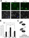
Bag1-L overexpression increases cell death of CGNs. (A and B) Primary cerebellar neurons were isolated and cultured in medium containing high K+, which promotes their survival. At day in vitro 6 (DIV6), CGNs were transfected with Bag1-L, Bag1-LΔBD, or empty vector together with GFP to mark transfected cells. Cell death was measured after 48 h (including ±12 h of K+ withdrawal, which induces apoptosis) and is shown as a percentage of total cells analyzed. Representative images of each condition indicating normal nuclei (white arrows) and apoptotic nuclei (blue arrows) are shown. An asterisk (P < 0.05) indicates statistical significance; n.s, not statistically significant. (C) A model illustrating the coactivation of apoptotic c-Jun/JNK signaling by Bag1-L in neurons.

Similar articles

Cited by

References

    1. Alberts, A. S., O. Geneste, and R. Treisman. 1998. Activation of SRF-regulated chromosomal templates by Rho-family GTPases requires a signal that also induces H4 hyperacetylation. Cell 92:475-487. - PubMed
    1. Angel, P., K. Hattori, T. Smeal, and M. Karin. 1988. The jun proto-oncogene is positively autoregulated by its product, Jun/AP1. Cell 55:875-885. - PubMed
    1. Barnes, J. D., N. J. Arhel, S. S. Lee, A. Sharp, M. Al-Okail, G. Packham, A. Hague, C. Paraskeva, and A. C. Williams. 2005. Nuclear BAG-1 expression inhibits apoptosis in colorectal adenoma-derived epithelial cells. Apoptosis 10:301-311. - PubMed
    1. Becker, E. B., J. Howell, Y. Kodama, P. A. Barker, and A. Bonni. 2004. Characterization of the c-Jun N-terminal kinase-BimEL signaling pathway in neuronal apoptosis. J. Neurosci. 24:8762-8770. - PMC - PubMed
    1. Behrens, A., M. Sibilia, and E. F. Wagner. 1999. Amino-terminal phosphorylation of c-Jun regulates stress-induced apoptosis and cellular proliferation. Nat. Genet. 21:326-329. - PubMed

Publication types

Substances