. 2010 Sep;51(9):2629-41.
doi: 10.1194/jlr.M005132.

Partition profile of the nicotinic acetylcholine receptor in lipid domains upon reconstitution

Affiliations

Partition profile of the nicotinic acetylcholine receptor in lipid domains upon reconstitution

Vicente Bermúdez et al. J Lipid Res. 2010 Sep.

Abstract

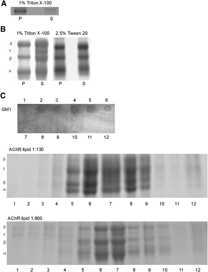
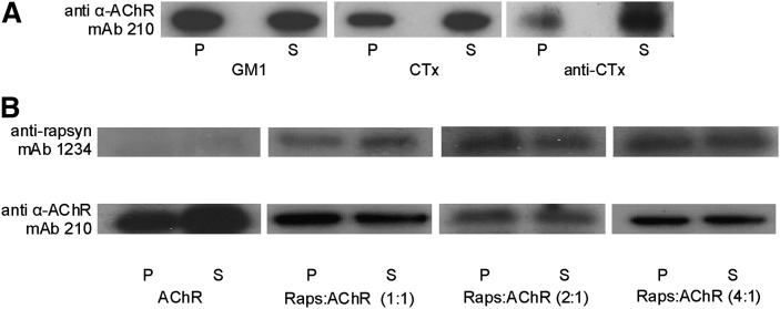
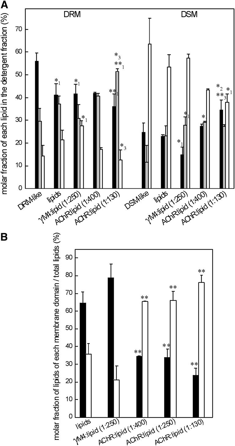
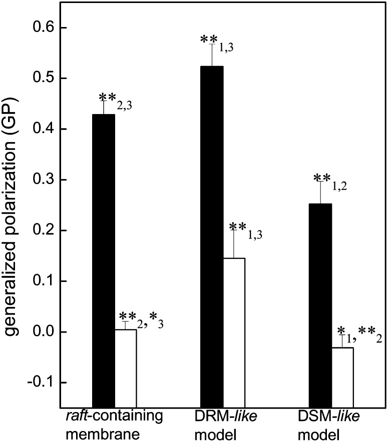
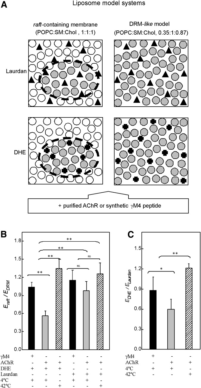
The nicotinic acetylcholine receptor (AChR) is in intimate contact with the lipids in its native membrane. Here we analyze the possibility that it is the intrinsic properties of the AChR that determine its partition into a given lipid domain. Torpedo AChR or a synthetic peptide corresponding to the AChR M4 segment (the one in closer contact with lipids) was reconstituted into "raft"-containing model membranes. The distribution of the AChR was assessed by Triton X-100 extraction in combination with fluorescence studies, and lipid analyses were performed on each sample. The influence of rapsyn, a peripheral protein involved in AChR aggregation, was studied. Raft-like domain aggregation was also studied using membranes containing the ganglioside GM1 followed by GM1 crosslinking. The gammaM4 peptide displays a marked preference for raft-like domains. In contrast, AChR alone or in the presence of rapsyn or ganglioside aggregation exhibits no such preference for raft-like domains, but it does cause a significant reduction in the total amount of these domains. The results indicate that the distribution of the AChR in lipid domains cannot be due exclusively to the intrinsic physicochemical properties of the protein and that there must be an external signal in native cell membranes that directs the AChR to a specific membrane domain.


Figures

Fig. 1.
Fig. 1.
Liposomes of (A) γM4 synthetic peptide in POPC:SM:Chol (1:1:1) and (B) purified AChR reconstituted in a POPC:SM:Chol (1:1:1) at a 1:130 AChR:lipid molar ratio were subjected to 1% Triton X-100 or 2.5% Tween 20 extraction at 4°C and subsequent high-speed centrifugation as described in “Materials and Methods.” DSM and DRM fractions were collected as supernatant (S) and pellet (P). C: Liposomes of purified AChR reconstituted in POPC: SM:Chol (1:1:1) at a 1:130 and 1:800 AChR:lipid molar ratios were subjected to 1% Triton X-100 extraction and subsequent sucrose density separation as described in “Materials and Methods.” A representative GM1 dot blot of fractions 1–12 is shown, indicating that the DRMs were located in fractions 3–6. Panels A–C are representative Coomassie blue-stained SDS-PAGE of at least three independent experiments. AChR, nicotinic acetylcholine receptor; Chol, cholesterol; DRM, detergent-resistant membrane; DSM, detergent-soluble membrane; POPC, palmitoyloleylphosphatydilcholine; SM, brain sphingomyelin.
Fig. 2.
Fig. 2.
A: Immunoblot with mAb 210 (monoclonal antibody against the α subunit of the T. californica AChR) of DRM (P) and DSM (S) fractions obtained from purified AChR reconstituted in POPC:SM:Chol containing GM1 (i) without further treatment, (ii) treated with cholera toxin B subunit (CTxB) for GM1 crosslinking, and (iii) treated with CTxB for GM1 crosslinking followed by anti-cholera toxin antibody (anti-CTx) for crosslinking of CTxB. B: Immunoblot of DSM (S) and DRM (P) fractions obtained from purified AChR reconstituted in a POPC:SM:Chol 1:1:1 system in the presence of a rapsyn-enriched extract in three different rapsyn:AChR molar stoichiometries (1:1, 2:1, and 4:1). Blots were probed with mAb 1234 (monoclonal antibody against rapsyn) and mAb 210 (monoclonal antibody against the α subunit of the T. californica AChR). Panels A and B are representative of at least three independent experiments. AChR, nicotinic acetylcholine receptor; Chol, cholesterol; CTx, cholera toxin; CTxB, cholera toxin B-subunit; DRM, detergent-resistant membrane; DSM, detergent-soluble membrane; GM1, ganglioside GM1; POPC, palmitoyloleylphosphatydilcholine; SM, brain sphingomyelin.
Fig. 3.
Fig. 3.
A: Molar fractions of Chol (filled bars), SM (gray bars), and PC (empty bars) in DRM and DSM fractions obtained from a DRM-like model and a DSM-like model (PC:SM:Chol 0.35:1:0.87 and 1:0.11:0.24 molar ratio, respectively, both without AChR) and from four different raft-containing membranes (POPC:SM:Chol, 1:1:1 molar ratio) in the absence of AChR (lipids), in the presence of γM4 peptide (1:250), and in the presence of different AChR:lipid molar ratios (1:400 and 1:130). Each bar corresponds to the average ± SD of at least three independent measurements. Statistically significant differences: *P < 0.05; **P < 0.01. Subscripts indicate the lipid system with which the comparison showed statistically significant differences: (1) DRM- or DSM-like system, (2) raft-containing membrane, and (3) γM4:lipid model. B: Molar fractions of the lipids of DRM (black columns) or DSM (white columns) with respect to the total lipid of different model membranes in the absence of AChR (lipids), in the presence of γM4 peptide (1:250), and in the presence of different AChR:lipid molar ratios (1:400, 1:250, and 1:130). Each bar corresponds to the average ± SD of at least three independent measurements. Statistically significant differences with respect to the lipid system without AChR: *P < 0.05; **P < 0.01. AChR, nicotinic acetylcholine receptor; Chol, cholesterol; DRM, detergent-resistant membrane; DSM, detergent-soluble membrane; POPC, palmitoyloleylphosphatydilcholine; SM, brain sphingomyelin.
Fig. 4.
Fig. 4.
Excitation GP of Laurdan of purified AChR reconstituted in a raft-containing membrane (POPC:SM:Chol, 1:1:1 molar ratio), a DRM-like model (PC:SM:Chol, 0.35:1:0.87 molar ratio), and a DSM-like model (PC:SM:Chol, 1:0.11:0.24 molar ratio) at 4°C and 42°C (black and white columns, respectively). Each column corresponds to the average ± SD of at least four independent measurements. Statistically significant differences between black columns or white columns: *P < 0.05; **P < 0.01. The subindices indicate the lipid system with which the comparison showed statistically significant differences: (1) raft-containing membrane, (2) DRM-like model, and (3) DSM-like model. Although it is not shown in the figure, the differences between the black and white columns of each lipid system are statistically significant (P < 0.01). AChR, nicotinic acetylcholine receptor; Chol, cholesterol; DRM, detergent-resistant membrane; DSM, detergent-soluble membrane; GP, generalized polarization; PC, phosphatydilcholine; POPC, palmitoyloleylphosphatydilcholine; SM, brain sphingomyelin.
Fig. 5.
Fig. 5.
A: Lipid model systems used in FRET studies: raft-containing membrane (POPC:SM:Chol, 1:1:1 molar ratio), where there is a coexistence of two different lipid domains (⊘, ○ for DRM and DSM-like domains, respectively), and DRM-like model (POPC:SM:Chol, 0.35:1:0.87 molar ratio) consisting of only one lipid domain (⊘). Laurdan fluorescent probe (▴), used as a control probe, shows no preferential partition into a particular domain. DHE (✚), used as a fluorescent Chol mimetic, labels DRM domains. FRET measurements were performed between the intrinsic fluorescence of the γM4 peptide or the AChR, donor molecules, and Laurdan or DHE as acceptor probes. B: Ratio of the FRET efficiencies between the intrinsic fluorescence of the purified AChR (or the γM4 peptide) as donor molecules and DHE (or Laurdan) as acceptor molecules obtained with the raft-containing membrane (Eraft) and with the DRM-like model (EDRM) at 4°C and 42°C. C: Ratio of the FRET efficiencies obtained between the intrinsic fluorescence of the purified AChR (or the γM4 peptide) as donor molecules and DHE (EDHE) and Laurdan (ELaurdan) as acceptor molecules in the raft-containing membrane at 4°C and 42°C. Each column corresponds to the average ± SD of at least four independent measurements. *, ** Statistically significant differences: *P < 0.05; **P < 0.01. NS (nonsignificant) differences: P < 0.05. AChR, nicotinic acetylcholine receptor; Chol, cholesterol; DHE, dehydroergosterol; DRM, detergent-resistant membrane; DSM, detergent-soluble membrane; E, energy transfer efficiency; FRET, Förster resonance energy transfer; POPC, palmitoyloleylphosphatidilcholine; SM, brain sphingomyelin.
Fig. 6.
Fig. 6.
Gibbs triangle showing DRM (solid) and DSM (open) compositions for all the systems tested in this work: DRM-like (▪), DSM-like (□), different raft-containing membranes (i) in the absence of AChR (•, ○), (ii) in the presence of γM4 peptide (1:250 ▴, △), and (iii) in the presence of different AChR:lipid molar ratios (1:400, ▾, ▽; and 1:130, ♦, ⋄). The DRM and DSM lipid compositions proposed in the work of Schroeder et al. (50) (◂, ◃) and McIntosh et al. (51) (▸, ▹) are indicated. Also shown are the lines that divide the phase diagram in the different pure phases (solid, s; liquid-ordered, lo; and liquid disordered, ld) and the different coexistence regions at 22°C (s + lo; ld + lo + s; ld + s; and ld + lo) proposed by Pokorny et al. (70). AChR, nicotinic acetylcholine receptor; BSM, brain sphingomyelin; Chol, cholesterol; DRM, detergent-resistant membrane; DSM, detergent-soluble membrane; POPC, palmitoyloleylphosphatydilcholine.

Similar articles

Cited by

References

    1. Barrantes F. J. 2004. Structural basis for lipid modulation of nicotinic acetylcholine receptor function. Brain Res. Brain Res. Rev. 47: 71–95. - PubMed
    1. Barrantes F. J. 2003. Modulation of nicotinic acetylcholine receptor function through the outer and middle rings of transmembrane domains. Curr. Opin. Drug Discov. Devel. 6: 620–632. - PubMed
    1. Sanes J. R., Lichtman J. W. 2001. Induction, assembly, maturation and maintenance of a postsynaptic apparatus. Nat. Rev. Neurosci. 2: 791–805. - PubMed
    1. Moransard M., Borges L. S., Willmann R., Marangi P. A., Brenner H. R., Ferns M. J., Fuhrer C. 2003. Agrin regulates rapsyn interaction with surface acetylcholine receptors, and this underlies cytoskeletal anchoring and clustering. J. Biol. Chem. 278: 7350–7359. - PubMed
    1. Borges L. S., Ferns M. 2001. Agrin-induced phosphorylation of the acetylcholine receptor regulates cytoskeletal anchoring and clustering. J. Cell Biol. 153: 1–12. - PMC - PubMed

Publication types

LinkOut - more resources