. 2010 Jul;120(7):2497-507.
doi: 10.1172/JCI42382.

Distinct roles for PTEN in prevention of T cell lymphoma and autoimmunity in mice

Affiliations

Distinct roles for PTEN in prevention of T cell lymphoma and autoimmunity in mice

Xiaohe Liu et al. J Clin Invest. 2010 Jul.

Abstract

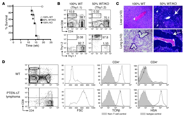
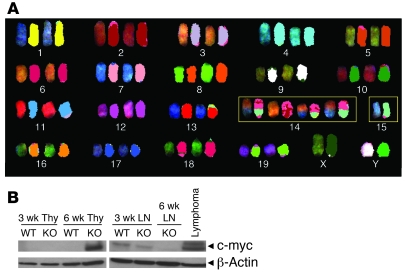
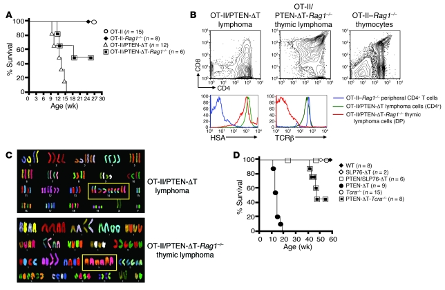
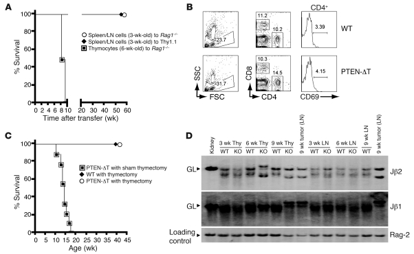
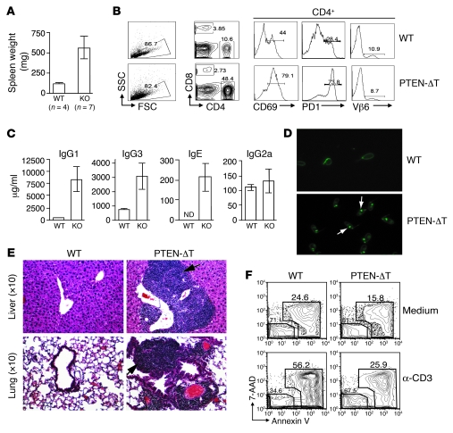
Mutations in the tumor-suppressor gene phosphatase and tensin homolog deleted on chromosome 10 (Pten) are associated with multiple cancers in humans, including T cell malignancies. Targeted deletion of Pten in T cells induces both a disseminated "mature phenotype" lymphoma and a lymphoproliferative autoimmune syndrome in mice. Here, we have shown that these two diseases are separable and mediated by T lineage cells of distinct developmental stages. Loss of PTEN was found to be a powerful driver of lymphomagenesis within the thymus characterized by overexpression of the c-myc oncogene. In an otherwise normal thymic environment, PTEN-deficient T cell lymphomas invariably harbored RAG-dependent reciprocal t(14:15) chromosomal translocations involving the T cell receptor alpha/delta locus and c-myc, and their survival and growth was TCR dependent, but Notch independent. However, lymphomas occurred even if TCR recombination was prevented, although these lymphomas were less mature, arose later in life, and, importantly, were dependent upon Notch pathways to upregulate c-myc expression. In contrast, using the complementary methods of early thymectomy and adoptive transfers, we found that PTEN-deficient mature T cells were unable to undergo malignant transformation but were sufficient for the development of autoimmunity. These data suggest multiple and distinct regulatory roles for PTEN in the molecular pathogenesis of lymphoma and autoimmunity.


Figures

Figure 1
Figure 1. Lymphomagenesis in PTEN-ΔT mice is T cell intrinsic.
(A) Bone marrow chimeras were prepared by transplanting bone marrow from WT mice (n = 4), PTEN-ΔT mice (n = 4), or a 50:50 mixture of the two (n = 8) into lethally irradiated WT congenic recipients. Survival of chimeric mice is measured in weeks after reconstitution. (B) Flow cytometric analyses of T cells in the spleen of representative chimeric mice from A and the contribution of WT (Thy1.1) and PTEN-ΔT (Thy1.2) bone marrow to the peripheral CD4 population. (C) H&E staining of liver and lung from representative chimeric mice 15 weeks after reconstitution. (D) Immunophenotype of lymphoma cells in PTEN-ΔT mice. Cells from lymphoid organs of 10- to 17-week-old PTEN-ΔT mice with CD4 single-positive lymphomas were stained with antibodies for TCRβ or HSA and analyzed by flow cytometry. Forward scatter (FSC) is an indicator of cell size. Data are representative of 5 experiments.
Figure 2
Figure 2. t(14;15) translocations and c-myc activation in lymphomas from PTEN-ΔT mice.
(A) Detection of t(14;15) by SKY analysis in metaphases from lymphoma cells in lymph nodes of diseased PTEN-ΔT mice (>9 wk). Note that distinct colors in a single chromosome are indicative of a translocation. Similar results were seen in lymphoma cells isolated from the spleen. Image shown is representative of total of 127 metaphases from 5 mice examined. (B) Western blot analysis of c-myc expression in tumor cells from the thymus (Thy) or lymph node (LN).
Figure 3
Figure 3. Lymphomagenesis in PTEN-ΔT mice defective in V(D)J gene recombination or TCR signaling.
(A) Survival of OT-II/PTEN-ΔT-Rag1–/– mice and littermate controls. (B) Lymphomas from diseased OT-II/PTEN-ΔT and OT-II/PTEN-ΔT-Rag1–/– mice and thymocytes from littermate controls were stained with the indicated antibodies and analyzed by flow cytometry. The data are representative of 3 independent experiments. DP, double-positive. (C) SKY analysis of lymphoma cells from OT-II/PTEN-ΔT-Rag1–/– mice and OT-II/PTEN-ΔT controls. Note that distinct colors in a single chromosome are indicative of a translocation. Image shown is representative of more than 91 metaphases from 3–5 mice per group. (D) Survival of PTEN/SLP76-ΔT and PTEN-ΔT-Tcra–/– mice.
Figure 4
Figure 4. Activated Notch signaling in t(14;15)-negative lymphoma cells.
(A) RT-PCR analysis of pre-Tα (pTα) expression in RNA isolated from the indicated cells. (B) Quantitative RT-PCR for Hes-1 and c-myc in WT thymocytes or indicated primary lymphoma cells normalized to Gapdh. (C) Flow cytometric analysis of CD25 expression in cell lines derived from the indicated primary lymphomas. (D) Growth suppression by GSI. The tumor cell lines derived from the indicated primary lymphoma cells were cultured with or without 1 μM GSI in a 24-well plate for 72 hours. Graph shows the number of trypan blue–negative cells per well. (E) Impaired proliferation, cell cycle arrest, and apoptosis in t(14;15)-negative tumor cell lines treated with GSI. (F) Western blot analysis of the activated form of Notch1 protein in the indicated cell lines. All experiments described in CF are representative of 6 or more different cell lines derived from at least 2 individual primary lymphomas.
Figure 5
Figure 5. Requirements for lymphomagenesis and autoimmunity in PTEN-ΔT mice.
(A) Rag1–/– mice or sublethally irradiated B6 Thy1.1 mice were adoptively transferred with 5 million thymocytes or peripheral T cells (purified from spleen or lymph node as indicated) from 3- or 6-week-old PTEN-ΔT Thy1.2 mice. (B) Lack of malignant transformation of T cells from 3- to 4-week-old PTEN-ΔT mice following adoptive transfer into Rag–/– mice at 100 days after transfer. (C) PTEN-ΔT or WT mice were thymectomized at 3.5 weeks of age and monitored for survival (n = 9 for each group). (D) Southern blot analysis of TCRβ rearrangement in genomic DNA of thymocytes. Paired thymocyte and LN T cell samples from mice of the indicated ages were obtained from the same individual mice. The sample of a lymphoma-bearing, 9-week-old PTEN-ΔT mouse represents tumor cells. Germline configurations of mouse kidney DNA shown for comparison. Data are representative of 3 premalignant mice of each group and 6 tumors. Rag-2 is shown as a loading control.
Figure 6
Figure 6. The development of autoimmunity in thymectomized PTEN-ΔT mice.
(A) Representative splenic weights from thymectomized mice at 9.5–11 months of age. (B) WT and PTEN-ΔT mice that received a thymectomy were sacrificed at 9.5 months of age. Data represent lymphocyte profiles from the spleen of representative aged, thymectomized mice. (C) Elevated immunoglobulin levels from sera of 9.5-month-old thymectomized WT and PTEN-ΔT mice from B measured by ELISA. (D) Detection of serum dsDNA autoantibodies in 9.5-month-old thymectomized WT and PTEN-ΔT mice. Sera from the indicated mice was used to detect the presence of anti-dsDNA antibodies. Kinetoplast staining (as indicated by the arrow) indicates a positive antibody response to dsDNA. Original magnification, ×10. (E) Representative H&E staining of liver and lung of thymectomized WT and PTEN-ΔT mice at 9.5 months of age. All experiments described in AE are representative of 4 mice of each group. (F) Defective AICD in PTEN-deficient CD4+ T cells. T cells were isolated from 3.5-week-old PTEN-ΔT and WT mice and cultured in the presence of α-CD3, α-CD28, and IL-2. Viable cells were fractionated and restimulated with α-CD3 for an additional 16 hours, and cell death was evaluated by flow cytometry with the indicated markers gated on CD4+ population. The data are representative of 2 independent experiments on T cells from a total of 6 mice of each genotype.

References

    1. Li J, et al. PTEN, a putative protein tyrosine phos­phatase gene mutated in human brain, breast, and prostate cancer. Science. 1997;275(5308):1943–1947. - PubMed
    1. Maehama T, Dixon JE. The tumor suppressor, PTEN/MMAC1, dephosphorylates the lipid second messenger, phosphatidylinositol 3,4,5-trisphosphate. J Biol Chem. 1998;273(22):13375–13378. - PubMed
    1. Engelman JA, Luo J, Cantley LC. The evolution of phosphatidylinositol 3-kinases as regulators of growth and metabolism. Nat Rev Genet. 2006;7(8):606–619. - PubMed
    1. Teng DH, et al. MMAC1/PTEN mutations in primary tumor specimens and tumor cell lines. Cancer Res. 1997;57(23):5221–5225. - PubMed
    1. Liaw D, et al. Germline mutations of the PTEN gene in Cowden disease, an inherited breast and thyroid cancer syndrome. Nat Genet. 1997;16(1):64–67. - PubMed

Publication types

MeSH terms

Substances