. 2010 Aug;30(8):1432-6.
doi: 10.1038/jcbfm.2010.73. Epub 2010 Jun 2.

Laser speckle contrast imaging of collateral blood flow during acute ischemic stroke

Affiliations

Laser speckle contrast imaging of collateral blood flow during acute ischemic stroke

Glenn A Armitage et al. J Cereb Blood Flow Metab. 2010 Aug.

Abstract

Collateral vasculature may provide an alternative route for blood flow to reach the ischemic tissue and partially maintain oxygen and nutrient support during ischemic stroke. However, much about the dynamics of stroke-induced collateralization remains unknown. In this study, we used laser speckle contrast imaging to map dynamic changes in collateral blood flow after middle cerebral artery occlusion in rats. We identified extensive anastomatic connections between the anterior and middle cerebral arteries that develop after vessel occlusion and persist for 24 hours. Augmenting blood flow through these persistent yet dynamic anastomatic connections may be an important but relatively unexplored avenue in stroke therapy.


Figures
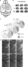
Figure 1
Figure 1
Surgical preparation, histology, and blood flow mapping in sham animals. (A) Imaging windows were placed 1 mm lateral from the bregma directly over the primary motor and somatosensory cortex for the forelimb and hindlimb. (B) Representative hematoxylin and eosin (H&E) staining showing representative sections from two animals after middle cerebral artery occlusion (MCAo) (top two rows) and a sham animal. Sections were chosen to approximately match the stereotaxic coordinates (Bregma±2.52 mm) in C. (Panel C) The largest and smallest infarcts induced by thromboembolic MCAo. Infarcts were identified on the basis of H&E staining in the brains extracted 24 hours after ischemic onset (coordinates and brain sections based on the atlas by Paxinos and Watson, 2007). (D) LSCI imaging showing a map of single vessel blood flow in the surface veins (V) and branches of the MCA in two sham-MCAo animals (top and bottom). Sham animals did not show any significant changes in blood flow or exhibit any anastomatic connections between the anterior cerebral artery (ACA) or MCA branches after sham-MCAo.
Figure 2
Figure 2
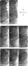
Mapping blood flow after focal stroke using laser speckle contrast imaging (LSCI). (A) LSCI image showing blood flow in the surface veins (V) and branches of the middle cerebral artery (MCA) over the hindlimb and forelimb sensorimotor cortex. It is noteworthy that blood flows through anastomatic connections between the distal segments of the anterior cerebral artery (ACA) and MCA immediately after MCA occlusion (MCAo) (arrows). (B) Left and middle panels show blood flow before and immediately after MCAo mapped with LSCI, whereas the right panel shows blood flow 24 hours after stroke. Spontaneous reperfusion occurred between imaging sessions and blood flow through anastomatic connections ceased by 24 hours (large arrows). Darker veins indicative of increased veinous blood flow from the territory of the MCA were observed after reperfusion (asterisks). (C) Left, middle, and right panels show LSCI blood flow maps before, after, and 24 hours after MCAo. Arrows indicate new patterns of collateral flow through anastomatic connections between the ACA and MCA that were apparent soon after MCAo and persisted at 24 hours after onset. The small arrow identifies an anastamose with blood flow 24 hours after MCAo that was not apparent immediately after onset. A, anterior; P, posterior; M, medial; L, lateral.

References

    1. Ayata C, Dunn AK, Gursoy-Ozdemir Y, Huang Z, Boas DA, Moskowitz MA. Laser speckle flowmetry for the study of cerebrovascular physiology in normal and ischemic mouse cortex. J Cereb Blood Flow Metab. 2004;24:744–755. - PubMed
    1. Bang OY, Saver JL, Buck BH, Alger JR, Starkman S, Ovbiagele B, Kim D, Jahan R, Duckwiler GR, Yoon SR, Vinuela F, Liebeskind DS. Impact of collateral flow on tissue fate in acute ischaemic stroke. J Neurol Neurosurg Psychiatry. 2008;79:625–629. - PMC - PubMed
    1. Duncan DD, Kirkpatrick SJ. Can laser speckle flowmetry be made a quantitative tool. J Opt Soc Am A Opt Image Sci Vis. 2008;25:2088–2094. - PMC - PubMed
    1. Dunn AK, Bolay H, Moskowitz MA, Boas DA. Dynamic imaging of cerebral blood flow using laser speckle. J Cereb Blood Flow Metab. 2001;21:195–201. - PubMed
    1. Green AR. Pharmacological approaches to acute ischemic stroke: reperfusion certainly, neuroprotection possibly. Br J Pharmacol. 2008;153:S325–S338. - PMC - PubMed

Publication types

MeSH terms