. 2008 Jul 15;3(1-2):9-20.
doi: 10.2478/v10053-008-0010-7.

Visual masking: past accomplishments, present status, future developments

Affiliations

Visual masking: past accomplishments, present status, future developments

Bruno G Breitmeyer. Adv Cogn Psychol. .

Abstract

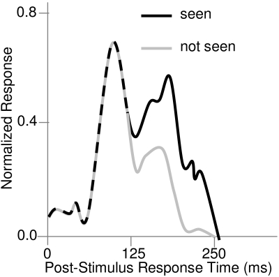
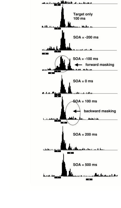
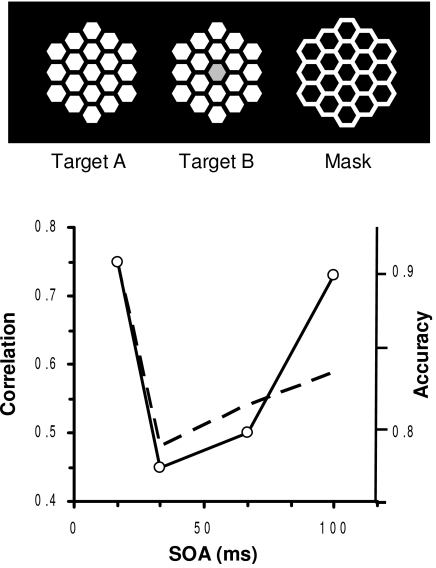
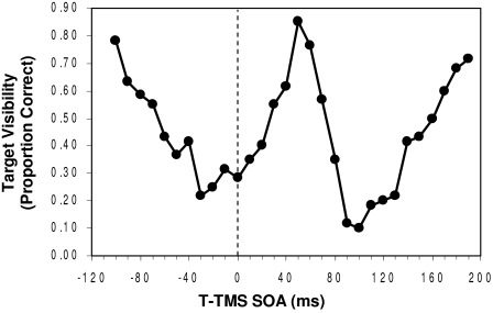
Visual masking, throughout its history, has been used as an investigative tool in exploring the temporal dynamics of visual perception, beginning with retinal processes and ending in cortical processes concerned with the conscious registration of stimuli. However, visual masking also has been a phenomenon deemed worthy of study in its own right. Most of the recent uses of visual masking have focused on the study of central processes, particularly those involved in feature, object and scene representations, in attentional control mechanisms, and in phenomenal awareness. In recent years our understanding of the phenomenon and cortical mechanisms of visual masking also has benefited from several brain imaging techniques and from a number of sophisticated and neurophysiologically plausible neural network models. Key issues and problems are discussed with the aim of guiding future empirical and theoretical research.

Keywords: masking; neural networks; nonconscious/conscious processing; object perception.


Figures

Figure 1.
Figure 1.
Post-stimulus multi-unit response magnitude functions obtained from V1 monkey neurons when a stimulus is perceived/ seen and when it is not perceived/seen. (Adapted from Lamme, Super, Landman, Roelfsema, & Spekreijse, 2000)
Figure 2.
Figure 2.
Multi-unit recordings from upper layers of area V1 of rhesus monkey. Note as indicated by dashed ovals a) optimal suppression of the early onset response component at a paracontrast SOA of -100 ms and b) optimal suppression of the later response component at a metcontrast SOA of 100 ms. (From Macknik & Livingstone, 1998)
Figure 3.
Figure 3.
Upper panel: “Honeycomb” target and mask stimuli. Lower panel: Correlation, derived from the fMRI results of the same observer, between activity in V1 level and the fusiform- gyrus (FG) level of cortical processing as a function of the SOA between the targets and the mask. (From Haynes, Driver & Rees, 2005)
Figure 4.
Figure 4.
Visibility (in proportion correct identification) of the target as a function of the onset asynchrony separating it from the TMS pulse. Negative SOAs: TMS precedes target; positive SOAs: TMS follows target. (Adapted from Corthout, Uttl, Ziemann et al., 1999).
Figure 5.
Figure 5.
(a) Comparison of a typical masking function obtained in our laboratory using a visual para- or metacontrast mask and a typical masking function obtained by Corthout, Uttl, Ziemann et al. (1999) using a TMS pulse as a mask. Negative and positive SOAs indicate that the masks were presented before and after the target, respectively. Results are not adjusted for retinocortical transmission delay. (b) Same as preceding but with results adjusted for a 60-ms delay of cortical M activity due to retinocortical transmission time (Baseler & Sutter, 1997). (From Breitmeyer, Ro, Öğmen, 2004)
Figure 6.
Figure 6.
Schematic of hypothetical metacontrast suppression of reentrant activation in the cortical parvocellular (P) pathways.
Figure 7.
Figure 7.
Metacontrast contour and surface-contrast suppression as a function of stimulus onset asynchrony (SOA). (Adapted after Breitmeyer et al., 2006)
Figure 8.
Figure 8.
Paracontrast contour suppression as a function of SOA. Note the two minima in target contour visibility at -200 and -10 ms. (Adapted after Breitmeyer et al., in press)
Figure 9.
Figure 9.
Target and mask visibilities (in proportion correct stimulus identification) under nonrivalrous (standard dichoptic) viewing of the target and the mask and under viewing in which the visibility of the mask is suppressed during binocular rivalry.

References

    1. Alpern M. Metacontrast. J. Opt. Soc. Am. 1953;43:648–657. - PubMed
    1. Andreassi J.L., De Simone J.J., Mellers B.W. Amplitude changes in the visual evoked potential with backward masking. Electroencephalogr. Clin. Neurophysiol. 1975;41:387–398. - PubMed
    1. Averbach E., Coriell A. S. Short-term memory in vision. Bell. Syst. Tech. J. 1961;40:309–328.
    1. Bachmann T. The process of perceptual retouch: Nonspecific afferent activation dynamics in explaining visual masking. Percept. & Psychophys. 1984;35:69–84. - PubMed
    1. Bachmann T. Psychophysiology of visual masking: The fine structure of conscious experience. Commack, NY: Nova Science Publishers; 1994.

LinkOut - more resources