. 2010 Jun;67(6):715-25.
doi: 10.1002/ana.21995.

Clinical progression in Parkinson disease and the neurobiology of axons

Affiliations

Clinical progression in Parkinson disease and the neurobiology of axons

Hsiao-Chun Cheng et al. Ann Neurol. 2010 Jun.

Abstract

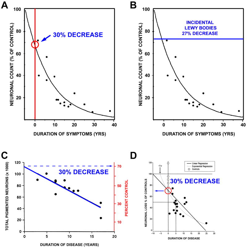
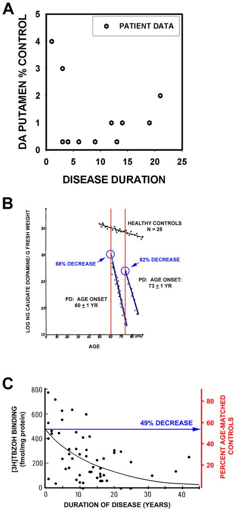
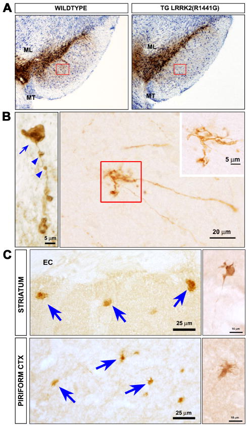
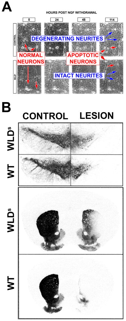
Despite tremendous growth in recent years in our knowledge of the molecular basis of Parkinson disease (PD) and the molecular pathways of cell injury and death, we remain without therapies that forestall disease progression. Although there are many possible explanations for this lack of success, one is that experimental therapeutics to date have not adequately focused on an important component of the disease process, that of axon degeneration. It remains unknown what neuronal compartment, either the soma or the axon, is involved at disease onset, although some have proposed that it is the axons and their terminals that take the initial brunt of injury. Nevertheless, this concept has not been formally incorporated into many of the current theories of disease pathogenesis, and it has not achieved a wide consensus. More importantly, in view of growing evidence that the molecular mechanisms of axon degeneration are separate and distinct from the canonical pathways of programmed cell death that mediate soma destruction, the possibility of early involvement of axons in PD has not been adequately emphasized as a rationale to explore the neurobiology of axons for novel therapeutic targets. We propose that ongoing degeneration of axons, not cell bodies, is the primary determinant of clinically apparent progression of disease, and that future experimental therapeutics intended to forestall disease progression will benefit from a new focus on the distinct mechanisms of axon degeneration.


Conflict of interest statement

Statement of Conflict: The authors have no financial interests to disclose

Figures

Fig. 1
Fig. 1
Estimates of loss of SN dopamine neurons at the time of PD symptom onset. (A) Fearnley and Lees examined the number of pigmented neurons in the SN in relation to duration of symptoms, and performed a regression analysis based on an exponential decline in their number. They estimated about a 30% total loss, adjusted for age. (B) In keeping with this estimate, they also found a sub-threshold loss of 27% of these neurons among individuals with incidental Lewy bodies. (C) A similar estimate can be derived from the data of Ma and colleagues, based on their dissector counts of pigmented SN neurons. A linear regression analysis of their data with extrapolation to time of disease onset yields an estimate of about a 30% loss. (D) Greffard and colleagues performed counts of neurons in the SNpc, and determined density per unit volume. By either a linear or a negative exponential best fit analysis, they estimated a 30% loss at the time of symptom onset.
Fig. 2
Fig. 2
Estimates of loss of striatal dopamine terminals markers at time of symptom onset. (A) A graphical representation of the data presented in the oft-cited Bernheimer study to support the statement that there is an 80% reduction at the time of disease onset. In the Bernheimer study, only 13 brains from patients with a diagnosis of PD were subjected to biochemical analysis. No regression analysis was performed. (B) Reiderer and Wuketich measured caudate dopamine content in two PD cohorts, one with an age of onset at 60 ± 1 years, and a second at 73 ± 1 years. Back extrapolation indicates a 68% and an 82% decrease, respectively, in caudate dopamine at the time of onset of disease in the two groups. (C) Scherman and colleagues analyzed [3H]TBZOH binding to the vesicular monoamine transporter in postmortem caudate nucleus of 54 PD patients. Polynomial regression analysis indicated a loss of 49% of binding sites at time of disease onset.
Fig. 3
Fig. 3
Axonopathy in hLRRK2(R1441G) BAC transgenic mice. (A) Immunoperoxidase staining for TH reveals no loss of SN dopamine neurons in the hLRRK2(R1441G) transgenics. (B) At the single axon level, staining for tyrosine hydroxylase (TH) reveals fragmentation (blue arrowheads), axonal spheroids (blue arrow), and dystrophic neurites at axon terminals (red square and inset). (C) Axonal abnormalities in the striatum and piriform cortex of the transgenic mice are also revealed by immunostaining for phosphorylated tau. Spheroids (blue arrows) and dystrophic neurites (side panels) similar to those visualized by TH staining, are observed. (Images adapted from Li et al43).
Fig. 4
Fig. 4
Studies of the WldS mutant mouse demonstrate that degeneration of neuron cell bodies and axons is mediated by distinct mechanisms. (A) Deckwerth and Johnson demonstrated that both the cell bodies and the neurites of wild type sympathetic ganglion neurons degenerate in culture after NGF withdrawal, but only cell bodies of WldS mice degenerate. (B) In a living mouse model Sajadi et al demonstrated that, remarkably, after medial forebrain bundle 6OHDA lesion, while there has been a substantial loss of neurons in mice of both genotypes, there is a substantial preservation of axons in WldS. The top panels show loss of dopamine neurons by immunostaining for TH, and the bottom panels show striatal dopaminergic innervation by immunostaining for the dopamine transporter.
Fig. 5
Fig. 5
Resistance of neuron cell bodies, but not axons, to degeneration in JNK null mice. (A) The intrastriatal 6OHDA neurotoxin model induces apoptosis in SN dopamine neurons in wildtype mice. A typical example of an apoptotic profile, with characteristic chromatin clumps, is demonstrated by thionin counterstain in the inset. The homozygous jnk2 and jnk3 single null mutations suppressed apoptosis by 95% and 98%, respectively, and the homozygous jnk2/3 double null mutations completely abrogated apoptosis. (B) The homozygous jnk2/3 double null mutations provided virtually complete protection of SN dopamine neurons. Among wildtype (WT) mice, there was a 63% loss of dopamine neurons, typical for this model, whereas among jnk2/3 nulls, there was only a 4% decrease. Low power photomicrographs of representative TH-immunostained SN sections from wildtype (top) and jnk2/3 nulls (bottom) following unilateral 6OHDA injection. (C) Homozygous jnk2/3 double null mice are not resistant to retrograde degeneration of nigrostriatal dopaminergic axons induced by intrastriatal 6OHDA. Following 6OHDA, there is a virtually complete loss of TH-positive fiber staining in homozygous jnk2/3 null mice, as in wildtype, throughout the striatum.

References

    1. Bergman H, Wichmann T, DeLong MR. Reversal of experimental parkinsonism by lesions of the subthalamic nucleus. Science. 1990;249:1436–1438. - PubMed
    1. Limousin P, Pollak P, Benazzouz A, et al. Effect of parkinsonian signs and symptoms of bilateral subthalamic nucleus stimulation. Lancet. 1995;345:91–95. - PubMed
    1. Shoulson I. Experimental therapeutics of neurodegenerative disorders: unmet needs. Science. 1998;282:1072–1074. - PubMed
    1. Olanow CW, Kieburtz K, Schapira AH. Why have we failed to achieve neuroprotection in Parkinson's disease? Ann Neurol. 2008;64 2:S101–S110. - PubMed
    1. Ellis RE, Yuan J, Horvitz HR. Mechanisms and functions of cell death. Annual Review of Cell Biology. 1991;7:663–698. - PubMed

Publication types