. 2010 Aug 2;7(4):1007-14.
doi: 10.1021/mp1001125.

Paclitaxel-loaded polymeric micelles modified with MCF-7 cell-specific phage protein: enhanced binding to target cancer cells and increased cytotoxicity

Affiliations

Paclitaxel-loaded polymeric micelles modified with MCF-7 cell-specific phage protein: enhanced binding to target cancer cells and increased cytotoxicity

Tao Wang et al. Mol Pharm. .

Abstract

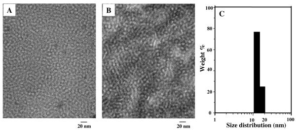
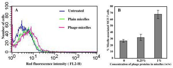
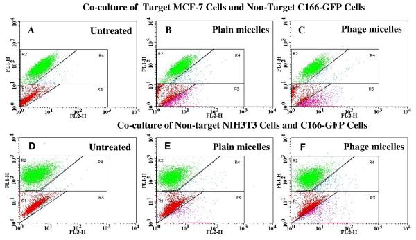
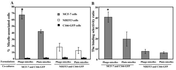
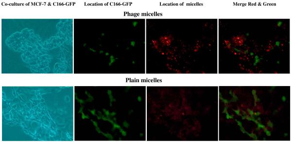
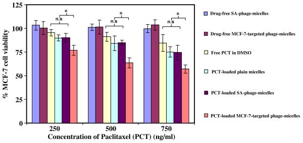
Polymeric micelles are used as pharmaceutical carriers to increase solubility and bioavailability of poorly water-soluble drugs. Different ligands are used to prepare targeted polymeric micelles. Earlier, we developed the method for use of specific landscape phage fusion coat proteins as targeted delivery ligands and demonstrated the efficiency of this approach with doxorubicin-loaded PEGylated liposomes. Here, we describe a MCF-7 cell-specific micellar formulation self-assembled from the mixture of the micelle-forming amphiphilic polyethylene glycol-phosphatidylethanolamine (PEG-PE) conjugate, MCF-7-specific landscape phage fusion coat protein, and the hydrophobic drug paclitaxel. These micelles demonstrated a very low cmc value and specific binding to target cells. Using an in vitro coculture model, FACS analysis, and fluorescence microscopy we showed that MCF-7 targeted phage-micelles preferentially bound to target cells compared to nontarget cells. As a result, targeted paclitaxel-loaded phage-micelles demonstrated a significantly higher cytotoxicity toward target MCF-7 cells than free drug or nontargeted micelle formulations, but failed to show such a differential toxicity toward nontarget C166 cells. Overall, cancer cell-specific phage proteins identified from phage display peptide libraries can serve as targeting ligands ("substitute antibody") for polymeric micelle-based pharmaceutical preparations.


Figures

Figure 1
Figure 1. Characterization of micellar formulations
A) TEM micrograph of paclitaxel-loaded phage PEG-PE micelles; B) TEM micrograph of drug-free plain PEG-PE micelles; C) Size distribution of phage micelles measured by dynamic light scattering.
Figure 2
Figure 2. FACS analysis of micellar uptake by MCF-7 cells indicated as fluorescence intensity of cell-associated rhodamine-labeled micelles
A) The histogram plot of the binding of rhodamine-labeled MCF-7-targeted phage-micelles and plain micelles to MCF-7 cells; B) Effect of concentration of phage proteins on binding affinity of micellar formulations to MCF-7 cells (mean ± SD, n=6).
Figure 3
Figure 3. FACS analysis of cell binding of micellar preparations in co-culture systems
Co-culture composed of target MCF-7 cells and non-target C166-GFP cells and control co-culture composed of non-target NIH3T3 cells and C166-GFP cells were incubated with either plain micelles or MCF-7-targeted phage-micelles for 1h, and micelle-associated cells ( as red fluorescence increase ) were analyzed using FACS. The dot plots were inserted into four regions (R1, R2, R3, and R4). FL1-H (green); FL2-H (red); A) Untreated co-culture of MCF-7 cells and C166-GFP cells. Red dots in R1 region show the location of untreated MCF-7 cells and green dots in R2 region show the location of untreated C166-GFP cells; B) Plain micelle-treated co-culture of MCF-7 cells and C166-GFP cells. Pink dots in R3 region show plain micelle-associated MCF-7 cells, and blue dots in R4 show plain micelle-associated C166-GFP cells; C) MCF-7-targeted phage-micelle-treated co-culture of MCF-7cells and C166-GFP cells. Pink dots in R3 region show phage-micelle-associated MCF-7 cells, and blue dots in R4 show phage-micelle-associated C166-GFP cells; D) Untreated control co-culture of NIH3T3 cells and C166-GFP. Red dots in R1 region show the location of untreated NIH3T3 cells, and green dots in R2 region show the location of untreated C166-GFP cells; E) Plain micelle-treated co-culture of NIH3T3 and C166-GFP cells. Pink dots in R3 region show plain micelle-associated NIH3T3 cells, and blue dots in R4 show plain micelle-associated C166-GFP cells; F) MCF-7-targeted phage-micelle-treated co-culture of NIH3T3and C166-GFP cells. Pink dots in R3 region show phage-micelle-associated NIH3T3 cells and blue dots in R4 show phage-micelle-associated C166-GFP cells.
Figure 4
Figure 4. Quantitative analysis of the binding of micellar preparations with cocultured target and non-target cells by FACS
A) Micelle-associated cells indicated as the intensity of cell population shifted to the region with higher red fluorescence; B) The binding selectivity ratios defined as the percent of micelle-associated MCF-7 cells or the percent of micelle-associated NIH3T3 cells divided by the percent of micelle-associated C166-GFP cells (* p <0.05, mean ± SD, n=6).
Figure 5
Figure 5. The binding of rhodamine-labeled MCF-7-targeted phage-micelles and plain micelles to co-culture MCF-7 and C166-GFP cells revealed by florescence microscopy
Green fluorescence is produced by C166-GFP cells; Red fluorescence labels MCF-7-targeted phage-micelles.
Figure 6
Figure 6
Cytotoxicity of micelle preparations toward MCF-7 cells after 72h treatment with MCF-7-targeted phage-micelles and various controls, including free PCT in DMSO, PCT-loaded plain micelles, irrelevant SA-phage-micelles, drug-free MCF-7-targeted phage-micelles and drug-free SA-phage-micelles (* p <0.05, mean ± SD, n=6).

Similar articles

Cited by

References

    1. Allen TM. Ligand-Targeted Therapeutics in Anticancer Therapy. Nat Rev Cancer. 2002;2:750–763. - PubMed
    1. Torchilin VP, Lukyanov AN, Gao Z, Papahadjopoulos-Sternberg B. Immunomicelles: Targeted Pharmaceutical Carriers for Poorly Soluble Drugs. Proc Natl Acad Sci U S A. 2003;100:6039–6044. - PMC - PubMed
    1. Salvatore G, Beers R, Margulies I, Kreitman RJ, Pastan I. Improved Cytotoxic Activity Toward Cell Lines and Fresh Leukemia Cells of a Mutant Anti-CD22 Immunotoxin Obtained by Antibody Phage Display. Clin Cancer Res. 2002;8:995–1002. - PubMed
    1. Park JW, Kirpotin DB, Hong K, Shalaby R, Shao Y, Nielsen UB, Marks JD, Papahadjopoulos D, Benz CC. Tumor Targeting Using Anti-Her2 Immunoliposomes. J Control Release. 2001;74:95–113. - PubMed
    1. Seymour LW, Ferry DR, Anderson D, Hesslewood S, Julyan PJ, Poyner R, Doran J, Young AM, Burtles S, Kerr DJ. Hepatic Drug Targeting: Phase I Evaluation of Polymer-Bound Doxorubicin. J Clin Oncol. 2002;20:1668–1676. - PubMed

Publication types