. 2010 Jun;173(6):748-59.
doi: 10.1667/RR1948.1.

Novel synthetic SOD/catalase mimetics can mitigate capillary endothelial cell apoptosis caused by ionizing radiation

Affiliations

Novel synthetic SOD/catalase mimetics can mitigate capillary endothelial cell apoptosis caused by ionizing radiation

Ekaterina Vorotnikova et al. Radiat Res. 2010 Jun.

Abstract

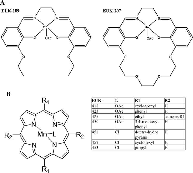
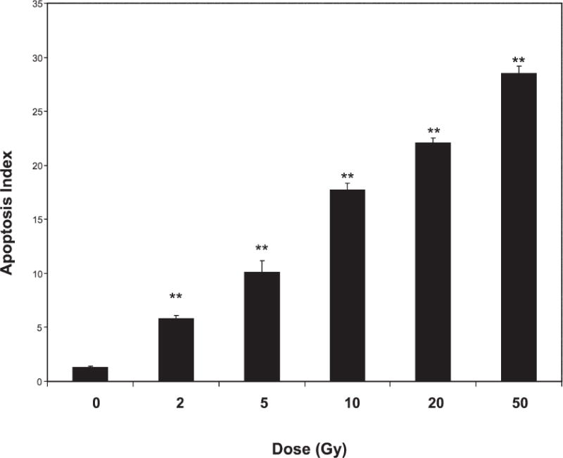
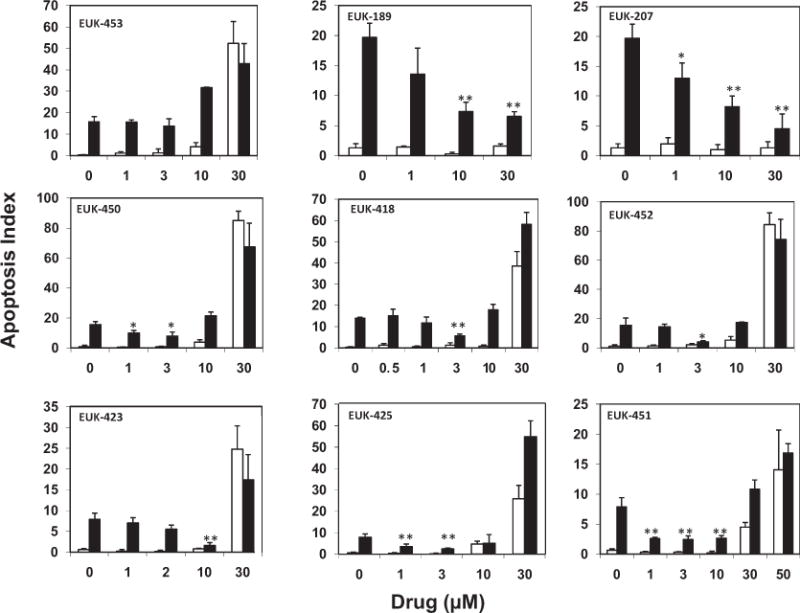
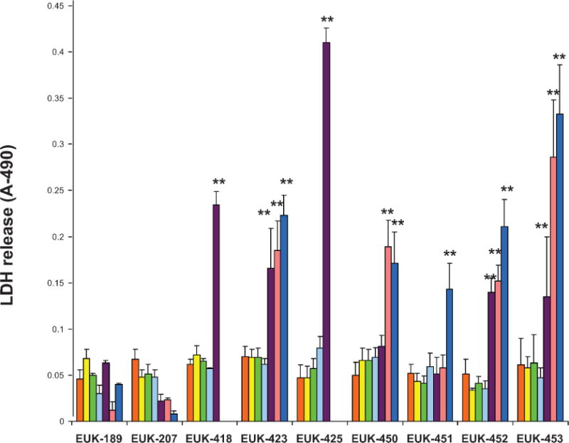
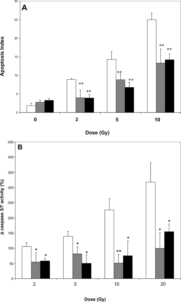
Numerous in vitro and in vivo studies have shown that the endothelial cells of the microvasculature of the lung and kidney are damaged by exposure to ionizing radiation, and this sustained endothelial cell injury is involved in the early and late radiation effects observed in these tissues. It is well accepted that ionizing radiation causes the generation of reactive oxygen species during exposure that results in damage to DNA and cellular organelles. It is more controversial, however, whether additional biochemical events or long-lived radicals occur and persist postirradiation that amplify and initiate new forms of cellular damage. Two families of Eukarion (EUK) compounds have been synthesized that possess superoxide dismutase (SOD), catalase and peroxidase activities. The Mn porphyrins are available orally whereas the salen Mn complexes are administered by injection. In the present study we have examined the ability of these SOD/catalase mimetics to prevent apoptosis of endothelial cells when administered 1 h postirradiation (mitigation). A range of salen Mn complex (EUK-189 and EUK-207) and Mn porphyrins (EUK-418, -423, -425, -450, -451, -452, -453) were used to treat endothelial cells 1 h after the cells received 2-20 Gy ionizing radiation in vitro. Two lead compounds, EUK-207 at a dose of 30 microM and EUK-451 at a dose of 10 microM, exhibited low toxicity and mitigated radiation-induced apoptosis. Future animal studies will test whether these compounds protect when administered after radiation exposure as would be done after a radiological accident or a terrorism event.


Figures

FIG. 1
FIG. 1
Panel A: The chemical structures of the salen Mn complexes EUK-189 and EUK-207. Panel B: The chemical structures of the Mn porphyrins compounds, the EUK-400 series.
FIG. 2
FIG. 2
Apoptosis of endothelial cells after X irradiation. Cells (passages 5–8) were sham-treated (0 Gy) or exposed to various doses of X rays; 6 h later, the number of cells undergoing apoptosis was determined. Differences between sham-treated and irradiated cells were compared using Student’s t test (**P < 0.001). Shown are pooled data from treatments performed in duplicate per experiment in three separate experiments. Error bars are SE.
FIG. 3
FIG. 3
Apoptosis of endothelial cells after sham (open bars) or 20 Gy irradiation (black bars) with or without various doses of EUK compounds administered 1 h postirradiation. Apoptosis indices were determined using cells at passage numbers 5–8 (top row), 9–11 (middle row) or 12–15 (bottom row). Statistically significant differences are shown for matched irradiated groups with and without drug. (Student’s t test, *P < 0.05; **P < 0.001). Bars are pooled data from treatments performed in duplicate per experiment. Error bars are SE.
FIG. 4
FIG. 4
Quantification of LDH released from endothelial cells (passages 5–8) treated with various doses of EUK compounds: 1 μM (yellow), 3 μM (green), 10 μM (light blue), 30 μM (purple), 50 μM (pink) and 100 μM (dark blue). Differences were compared between groups without drug (orange bars) compared to drug treatment using Student’s t tests (*P < 0.05; **P < 0.001). Error bars are SE.
FIG. 5
FIG. 5
Panel A: Effects of EUK compounds in preventing apoptosis of endothelial cells (passages 5–8) when applied 1 h after cells were exposed to radiation as determined by enumerating apoptotic bodies. Gray bars: 10 μM EUK-451; black bars: 30 μM EUK-207; open bars: no drug; **P < 0.001. Panel B: Effects of EUK 451 (gray bars) and EUK 271 (black bars) when applied to endothelial cells (passages 5–8) 1 h after cells were exposed to radiation (no drug, open bars) as detected by quantification of caspase 3/7. Bars represent changes in expression of executioner caspases in irradiated cultures from levels in sham cultures. *P < 0.05; **P < 0.001. Error bars are SE.

Similar articles

Cited by

References

    1. Nair CKK, Parida DK, Nomura T. Radioprotectors in radiotherapy. J Radiat Res. 2001;42:21–37. - PubMed
    1. Kerr JF, Wyllie AH, Currie AR. Apoptosis: a basic biological phenomenon with wide-ranging implications in tissue kinetics. Br J Cancer. 1972;26:239–257. - PMC - PubMed
    1. Wyllie AH, Kerr JFR, Currie AR. Cell death: the significance of apoptosis. Int Rev Cytol. 1990;68:251–306. - PubMed
    1. Edinger AL, Thompson CB. Death by design: apoptosis, necrosis and autophagy. Curr Opin Cell Biol. 2004;16:663–669. - PubMed
    1. Fink SL, Cookson BT. Apoptosis, pyroptosis, and necrosis: mechanistic description of dead and dying eukaryotic cells. Infect Immun. 2005;73:1907–1916. - PMC - PubMed

Publication types

MeSH terms