Review
. 2011 Feb 1;14(3):505-17.
doi: 10.1089/ars.2010.3331. Epub 2010 Aug 17.

Hypothalamic nitric oxide in hypoglycemia detection and counterregulation: a two-edged sword

Affiliations
Review

Hypothalamic nitric oxide in hypoglycemia detection and counterregulation: a two-edged sword

Xavier Fioramonti et al. Antioxid Redox Signal. .

Abstract

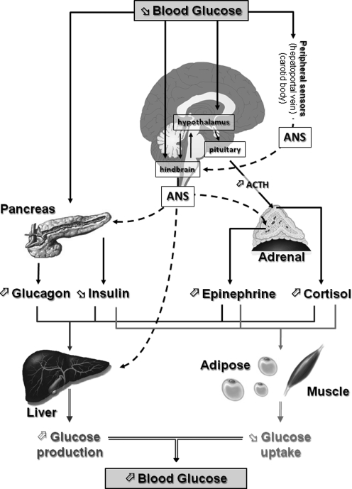
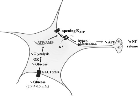
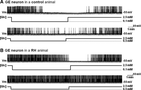
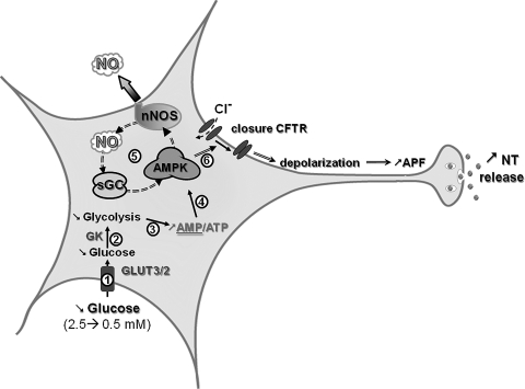
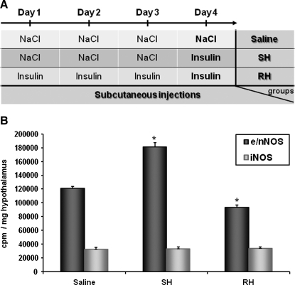
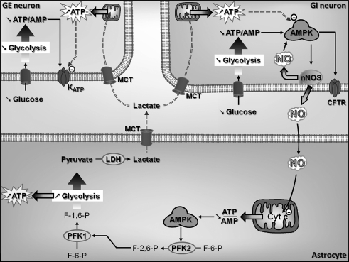
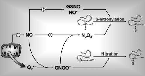
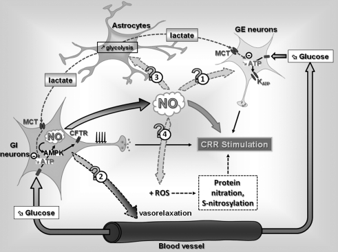
Hypoglycemia is the main complication for patients with type 1 diabetes mellitus receiving intensive insulin therapy. In addition to the obvious deleterious effects of acute hypoglycemia on brain function, recurrent episodes of hypoglycemia (RH) have an even more insidious effect. RH impairs the ability of the brain to detect and initiate an appropriate counterregulatory response (CRR) to restore euglycemia in response to subsequent hypoglycemia. Knowledge of mechanisms involved in hypoglycemia detection and counterregulation has significantly improved over the past 20 years. Glucose sensitive neurons (GSNs) in the ventromedial hypothalamus (VMH) may play a key role in the CRR. VMH nitric oxide (NO) production has recently been shown to be critical for both the CRR and glucose sensing by glucose-inhibited neurons. Interestingly, downstream effects of NO may also contribute to the impaired CRR after RH. In this review, we will discuss current literature regarding the molecular mechanisms by which VMH GSNs sense glucose. Putative roles of GSNs in the detection and initiation of the CRR will then be described. Finally, hypothetical mechanisms by which VMH NO production may both facilitate and subsequently impair the CRR will be discussed.


Figures

FIG. 1.
FIG. 1.
Neuroendocrine pathways involved in the CRR to hypoglycemia. Decreased blood glucose is detected by central (hypothalamus and hindbrain) and peripheral (pancreas, hepatoportal vein, and carotid body) glucose sensors. Together, these glucose sensors coordinate physiological responses that raise blood glucose levels. The initial response to hypoglycemia involves activation of the ANS, inhibition of insulin secretion, and stimulation of pituitary adrenocorticotropic hormone (ACTH) secretion. Activation of the autonomic nervous system increases glucagon and epinephrine secretion from the pancreas and adrenal medulla, respectively. ACTH stimulates cortisol release from the adrenal cortex. Increased glucagon, epinephrine, and cortisol together with decreased insulin stimulate hepatic glucose production and decrease adipose and muscle glucose uptake. The net result of the neuroendocrine CRR to hypoglycemia is to increase blood glucose levels and restore euglycemia. ANS, autonomic nervous system; CRR, counterregulatory response.
FIG. 2.
FIG. 2.
Molecular mechanisms by which VMN GE sense glucose. Decreased extracellular glucose leads to decreased glycolytic flux and decreases the ATP/AMP ratio. Decreased intracellular ATP level opens KATP channels that hyperpolarize the cell leading to decreased APF and NT release. APF, action potential frequency; GE, glucose-excited; KATP, ATP-sensitive K+; NT, neurotransmitter; VMN, ventromedial nucleus.
FIG. 3.
FIG. 3.
The response of VMN GE neurons to decreased glucose is impaired after RH. Continuous whole cell current clamp recordings of VMN GE neurons in brain slices from control rats (saline-injected; A) or animals injected daily for 3 consecutive days with insulin subcutaneously (RH protocol; B). (A) Decreased extracellular glucose from 2.5 to 0.1 mM (top trace) or from 2.5 to 0.5 mM (bottom trace) decreased membrane potential, APF, and input resistance in this VMN GE neuron from a control rat in a concentration-dependent manner. (B) In contrast, membrane potential, APF, and input resistance were only reduced in response to decreased glucose from 2.5 to 0.1 mM (top trace) but not to decreased glucose from 2.5 to 0.5 mM (bottom trace) in this VMN GE neuron from a rat exposed to RH. Changes in glucose concentration ([Glc]) are shown below each trace. Resting membrane potential (Vm) is represented by the dotted line and mentioned on the right of each trace. The downward deflections represent the membrane voltage response to a constant hyperpolarizing pulse (−20 pA) and were used to calculate input resistance. RH, recurrent hypoglycemia.
FIG. 4.
FIG. 4.
Molecular mechanisms by which VMN GI neurons sense glucose. Decreased extracellular glucose leads to decreased glycolytic flux and increases the AMP/ATP ratio ((1)–(3)). Increased AMP levels activate the AMPK ((4)). AMPK activation phosphorylates and activates nNOS increasing NO production. NO binds to its receptor sGC, which amplifies AMPK activation ((5)). This amplification of AMPK activity closes the CFTR chloride channel ((6)). CFTR closure depolarizes the GI neuron, leading to increased APF and NT release. AMPK, AMP-activated kinase; CFTR, cystic fibrosis transmembrane regulator; GI, glucose-inhibited; nNOS, neuronal nitric oxide synthase; sGC, soluble guanylate cyclase.
FIG. 5.
FIG. 5.
RH impairs insulin-induced increased NOS activity. (A) Experimental protocol of RH. (B) Ventral hypothalamic constitutive (e/nNOS) or iNOS NOS activity measured 60 min postinjection from rats injected subcutaneously with NaCl (saline) or insulin (4 U/kg) on the 4th day after 3 consecutive days of saline (SH) or insulin (RH) treatment. *p < 0.05 versus saline group (one-way ANOVA). iNOS, inducible NOS; SH, single hypoglycemia.
FIG. 6.
FIG. 6.
Hypothetical role of NO production by VMN GI neurons in the astrocyte-neuron lactate shuttle. Decreased extracellular glucose decreases GI neuronal intracellular ATP/AMP ratio and increases NO production through an AMPK-dependent mechanism. Full AMPK activation closes CFTR channels and increases GI neurons APF. Decreased extracellular glucose also decreases ATP/AMP ratio in GE neurons. Decreased ATP opens KATP channels and decreases GE neurons membrane potential and APF. NO produced by VMN GI neurons could diffuse to adjacent astrocytes and inhibit mitochondrial Cyt c and consequently mitochondrial respiration. Decreased mitochondrial respiration decreases astrocytic ATP levels, leading to AMPK activation. AMPK stimulates F-2,6-P formation via activation of PFK2. Increased F-2,6-P activates PFK1, which increases formation of F-1,6-P and consequently increases glycolytic flux. Increased glycolytic rate increases formation of anaerobic ATP and pyruvate. Pyruvate is converted lactate via LDH. Lactate may be transported to adjacent GI and GE neurons via MCT. In GI and GE neurons, lactate would increase intracellular ATP level, thus decreasing the response of GI and GE neurons to subsequent glucose decreases. Cyt c, cytochrome c; F-1,6-P, fructose-1,6-bisphosphate; F-2,6-P, fructose-2,6-bisphosphate; LDH, lactate dehydrogenase; MCT, monocarboxylate transporter; PFK, phospho-fructo-kinase.
FIG. 7.
FIG. 7.
Role of NO in protein posttranslational modification. Increased NO production can alter protein structure and ultimately function by nitration ((1)) and/or S-nitrosylation ((2), (3)). (1) NO can inhibit mitochondrial cytochrome c and stimulate generation of anion superoxide (O2•−). Interaction between NO and O2•− forms peroxynitrite (ONOO). ONOO can react with a free Tyr on proteins to form nitrated Tyr though a reaction called tyrosine nitration. (2) ONOO can also react with another molecule of NO to form dinitrogen trioxide (N2O3). N2O3 can interact with free Cys thiols on proteins to form S-nitro-thiol groups in a reaction called S-nitrosylation. (3) In an oxidative environment, increased NO level can also increase formation of GSNO and/or reactive NO (NO). GSNO and NO can also interact with free Cys to induce S-nitrosylation. Cys, cysteine; GSNO, nitroso-glutathione; Tyr, tyrosine.
FIG. 8.
FIG. 8.
Hypothetical mechanisms by which NO production by VMN GI neurons modulates the CRR to hypoglycemia. A decreased blood glucose level decreases the intracellular ATP level and stimulates NO production in VMN GI neurons through an AMPK-dependent mechanism, leading to increased APF. In VMN GE neurons, decreased blood glucose decreases the intracellular ATP level, leading to KATP opening and decreased APF. In addition to its role in GI neuronal glucose sensing and CRR initiation, NO produced by GI neurons could diffuse to adjacent cells, and (1) modulate GE neurons glucose sensitivity, (2) increase local blood flow, and/or (3) increase glycolytic flux in surrounding astrocytes. Increased astrocytic glycolysis would increase lactate production and transport to surrounding GI and GE neurons altering their glucose sensitivity. Finally ((4)), interaction between NO and ROS production could increase the level of protein nitration and/or S-nitrosylation and affect the CRR to subsequent hypoglycemia. ROS, reactive oxygen species.

Similar articles

Cited by

References

    1. The effect of intensive treatment of diabetes on the development and progression of long-term complications in insulin-dependent diabetes mellitus. The Diabetes Control and Complications Trial Research Group. N Eng J Med. 1993;329:977–986. - PubMed
    1. Almeida A. Moncada S. Bolanos JP. Nitric oxide switches on glycolysis through the AMP protein kinase and 6-phosphofructo-2-kinase pathway. Nat Cell Biol. 2004;6:45–51. - PubMed
    1. Alquier T. Kawashima J. Tsuji Y. Kahn BB. Role of hypothalamic Amp kinase in the impaired counterregulatory response induced by repetitive neuroglucopenia. Endocrinology. 2006;148:1367–1375. - PubMed
    1. Alquier T. Leloup C. Arnaud E. Magnan C. Penicaud L. Altered Glut4 mRNA levels in specific brain areas of hyperglycemic- hyperinsulinemic rats. Neurosci Lett. 2001;308:75–78. - PubMed
    1. Anand BK. Chhina GS. Sharma KN. Dua S. Singh B. Activity of single neurons in the hypothalamic feeding centers: effect of glucose. Am J Physiol. 1964;207:1146–1154. - PubMed

Publication types

MeSH terms