. 2010 Nov;131(3):318-30.
doi: 10.1111/j.1365-2567.2010.03309.x.

LAMP-2-deficient human B cells exhibit altered MHC class II presentation of exogenous antigens

Affiliations

LAMP-2-deficient human B cells exhibit altered MHC class II presentation of exogenous antigens

Victoria L Crotzer et al. Immunology. 2010 Nov.

Abstract

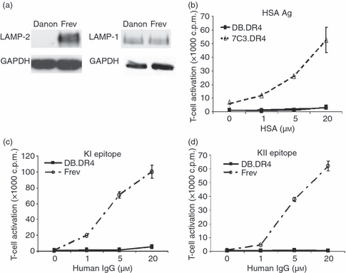
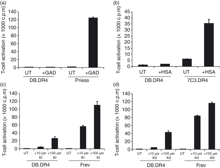
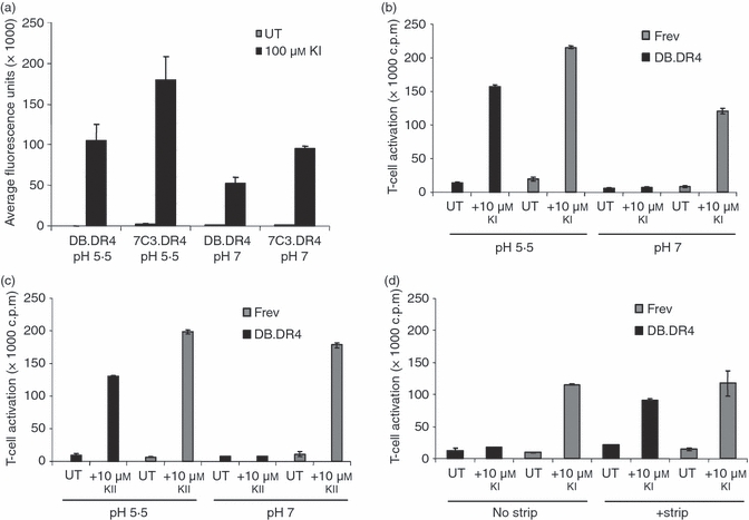
Major histocompatibility complex (MHC) class II molecules present antigenic peptides derived from engulfed exogenous proteins to CD4(+) T cells. Exogenous antigens are processed in mature endosomes and lysosomes where acidic proteases reside and peptide-binding to class II alleles is favoured. Hence, maintenance of the microenvironment within these organelles is probably central to efficient MHC class II-mediated antigen presentation. Lysosome-associated membrane proteins such as LAMP-2 reside in mature endosomes and lysosomes, yet their role in exogenous antigen presentation pathways remains untested. In this study, human B cells lacking LAMP-2 were examined for changes in MHC class II-restricted antigen presentation. MHC class II presentation of exogenous antigen and peptides to CD4(+) T cells was impaired in the LAMP-2-deficient B cells. Peptide-binding to MHC class II on LAMP-2-deficient B cells was reduced at physiological pH compared with wild-type cells. However, peptide-binding and class II-restricted antigen presentation were restored by incubation of LAMP-2-negative B cells at acidic pH, suggesting that efficient loading of exogenous epitopes by MHC class II molecules is dependent upon LAMP-2 expression in B cells. Interestingly, class II presentation of an epitope derived from an endogenous transmembrane protein was detected using LAMP-2-deficient B cells. Consequently, LAMP-2 may control the repertoire of peptides displayed by MHC class II molecules on B cells and influence the balance between endogenous and exogenous antigen presentation.


Figures

Figure 1
Figure 1
Exogenous antigen presentation is defective in LAMP-2-deficient Danon B-LCL. (a) Cell lysates were prepared from Danon or wild-type Frev B-LCL, the proteins were resolved by gel electrophoresis, and immunoblotted with an antibody to LAMP-2 or LAMP-1. The blot was stripped and reprobed with an antibody to GAPDH. Data are representative of at least two independent experiments. (b) LAMP-2-deficient DB.DR4 or wild-type 7C3.DR4 cells were incubated with various concentrations of human serum albumin antigen overnight and then cultured with HSA64–76-specific T cells to measure MHC class II presentation. (c, d) DB.DR4 or wild-type Frev cells were incubated with various concentrations of human IgG antigen overnight and then cultured with either κI188–203-specific T cells (c) or κII145–159-specific T cells (d) to measure MHC class II presentation. Neither DB.DR4 nor Frev expressed endogenous IgG κ light chain. Data are representative of three independent experiments, and the error bars indicate the mean T-cell activation ± the standard deviation. Note that the size of the symbols at some points masks the error bars.
Figure 2
Figure 2
Comparable expression of MHC class II messenger RNA and protein in Danon and wild-type B-LCL. (a) Total RNA was extracted from various wild-type or Danon B-LCL, complementary DNA was synthesized, and quantitative reverse transcription–polymerase chain reaction analysis performed using primers specific for HLA-DRα chain or for GAPDH as a control. Data are representative of the fold difference in messenger RNA expression observed in three independent experiments. (b) Wild-type Frev or LAMP-2-deficient DB.DR4 cells were incubated with L243 antibody to detect total surface HLA-DR and then stained with a fluorescein isothiocyanate-conjugated F(ab′)2 fragment of goat anti-mouse IgG secondary antibody. The mean fluorescence intensity (MFI) as measured by flow cytometry indicates the level of surface HLA-DR, and data are the average MFI of three independent experiments. (c) Cell lysates were prepared from LAMP-2-deficient DB.DR4 or wild-type Frev B-LCL, the proteins resolved by gel electrophoresis under non-reducing conditions to preserve MHC class II dimers, and immunoblotted with an antibody to HLA-DRα chain. Data are representative of at least five independent experiments. The ratios of HLA-DRαβ dimers to GAPDH as a loading control were 1·5 and 1·3 for DB.DR4 and Frev, respectively. (d) Various wild-type or Danon B-LCL were first permeabilized and then incubated with L243 or W6/32 antibody to detect total intracellular HLA-DR or MHC class I molecules, respectively. Cells were then stained with a phycoerythrin-conjugated F(ab′)2 fragment of rabbit anti-mouse immunoglobulin secondary antibody. The MFI as measured by flow cytometry indicates the levels of total surface or intracellular MHC class I or class II molecules. Data are the average MFI of three independent experiments. (e) Wild-type Priess and Frev expressing endogenous HLA-DR4 and wild-type 7C3.DR4 or LAMP-2-deficient DB.DR4 transfectants were incubated with 3.5.9-13F10 antibody to detect surface HLA-DR4β chains and then stained with a Cy2-conjugated F(ab′)2 fragment of donkey anti-rat IgG secondary antibody. The MFI as measured by flow cytometry indicates the level of surface HLA-DR4, and data are representative of more than three independent experiments. (f) Various wild-type or Danon B-LCL were first permeabilized and then incubated with MaP.DM1 (top panel) or a monoclonal antibody to HLA-DO (bottom panel) to detect total intracellular HLA-DM or HLA-DO molecules, respectively. Cells were then stained with a phycoerythrin-conjugated F(ab′)2 fragment of rabbit anti-mouse immunoglobulin secondary antibody. The MFI as measured by flow cytometry indicates the levels of total intracellular HLA-DM or HLA-DO molecules. Data for HLA-DM staining are the average MFI of three independent experiments while the data for HLA-DO staining are a representative experiment.
Figure 3
Figure 3
Endocytosis and MHC class II distribution in Danon and wild-type B-LCL. (a) Wild-type 7C3.DR4 or LAMP-2-deficient DB.DR4 B-LCL were incubated with fluorescein isothiocyanate (FITC) -albumin for 0 or 120 min at 37° and uptake was measured by flow cytometry. Data are representative of two independent experiments. (b) Wild-type Frev or LAMP-2-deficient DB.DR4 B-LCL were incubated with LysoTracker Red at 37° overnight to label acidic organelles, permeabilized, and then incubated with FITC-conjugated L423 antibody to detect HLA-DR. The results are representative of three independent experiments. (c) Wild-type Frev or LAMP-2-deficient DB.DR4 B-LCL were first permeabilized and then incubated with FITC-conjugated L423 antibody to detect HLA-DR and AlexaFluor647-conjugated-LAMP-1 Ab to detect LAMP-1. The results are representative of two independent experiments.
Figure 4
Figure 4
Efficient presentation of an endogenous antigen by Danon B-LCL. Variable numbers of DB.DR4 or 7C3.DR4 cells were cultured with HLA-A52–70-specific, HLA-DR4-restricted T cells to measure MHC class II presentation of endogenous HLA-A. As a control, the HLA-DR4 transfected murine B cell CH27.DR4 was left untreated or incubated with 10 μm HLA-A52–70 peptide and then cultured with HLA-A52–70-specific, HLA-DR4-restricted T cells. Data are representative of three independent experiments, and the error bars indicate the mean T-cell activation ± the standard deviation.
Figure 5
Figure 5
LAMP-2-deficient DB.DR4 cells fail to efficiently present exogenous peptides to CD4+ T cells. (a) LAMP-2-deficient DB.DR4 or wild-type Priess cells were left untreated (UT) or incubated with 10 μm exogenous GAD273–285 peptide for 4 hr and then cultured with GAD273–285-specific T cells to measure MHC class II presentation. (b) DB.DR4 or wild-type 7C3.DR4 cells were left untreated (UT) or incubated with 10 μm exogenous HSA64–76 peptide for 4 hr and then cultured with HSA64–76-specific T cells to measure MHC class II presentation. (c, d) DB.DR4 or wild-type Frev cells were left untreated (UT) or incubated with 10 μm or 100 μm exogenous κI188–203 peptide (c) or κII145–159 peptide (d) for 4 hr and then cultured with κI188–203-specific or κII145–159-specific T cells, respectively, to measure MHC class II presentation. Data in (a–d) are representative of three independent experiments, and the error bars indicate the mean T-cell activation ± the standard deviation.
Figure 6
Figure 6
Acidic pH restores exogenous peptide presentation by LAMP-2-deficient DB.DR4. (a) LAMP-2-deficient DB.DR4 or wild-type 7C3.DR4 cells were left untreated (UT) or incubated overnight with 100 μm biotinylated κI188–203 peptide. Lysates were prepared and added to plates coated with an anti-HLA-DR4 antibody to capture HLA-DR4 molecules in the lysates. The binding of biotinylated κI188–203 peptide to captured HLA-DR4 was measured using europium-strepavidin. The results are the average of three independent experiments, and the error bars indicate the standard deviation ± between the experiments. (b, c) DB.DR4 or wild-type Frev cells were left untreated (UT) or incubated with 10 μm exogenous κI188–203 peptide (b) or 10 μm exogenous κII145–159 peptide (c) in either cell culture medium (pH 7) or in sodium phosphate/citric acid buffer (pH 5·5) for 4 hr and then cultured with κI188–203-specific or κII145–159-specific T cells, respectively, to measure MHC class II presentation. Data in (b, c) are representative of three independent experiments, and the error bars indicate the mean T-cell activation ± the standard deviation. (d) DB.DR4 or Frev cells were left untreated (UT) or treated with a NaCl/citric acid buffer to strip surface MHC class II before incubation with 10 μm exogenous κI188–203 peptide for 4 hr. Cells were then cultured with κI188–203-specific T cells to measure MHC class II presentation. Data are representative of two independent experiments, and the error bars indicate the mean T-cell activation ± the standard deviation.

Similar articles

Cited by

References

    1. Watts C. The exogenous pathway for antigen presentation on major histocompatibility complex class II and CD1 molecules. Nat Immunol. 2004;5:685–92. - PubMed
    1. Cresswell P. Invariant chain structure and MHC class II function. Cell. 1996;84:505–7. - PubMed
    1. Sant AJ, Miller J. MHC class II antigen processing: biology of invariant chain. Curr Opin Immunol. 1994;6:57–63. - PubMed
    1. Maric MA, Taylor MD, Blum JS. Endosomal aspartic proteinases are required for invariant-chain processing. Proc Natl Acad Sci U S A. 1994;91:2171–5. - PMC - PubMed
    1. Riese RJ, Wolf PR, Bromme D, Natkin LR, Villadangos JA, Ploegh HL, Chapman HA. Essential role for cathepsin S in MHC class II-associated invariant chain processing and peptide loading. Immunity. 1996;4:357–66. - PubMed

Publication types

MeSH terms

Substances