. 2010 Nov;131(3):357-70.
doi: 10.1111/j.1365-2567.2010.03307.x.

Stromal cells differentially regulate neutrophil and lymphocyte recruitment through the endothelium

Affiliations

Stromal cells differentially regulate neutrophil and lymphocyte recruitment through the endothelium

Helen M McGettrick et al. Immunology. 2010 Nov.

Abstract

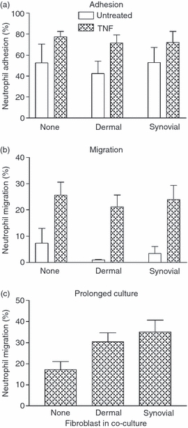
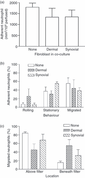
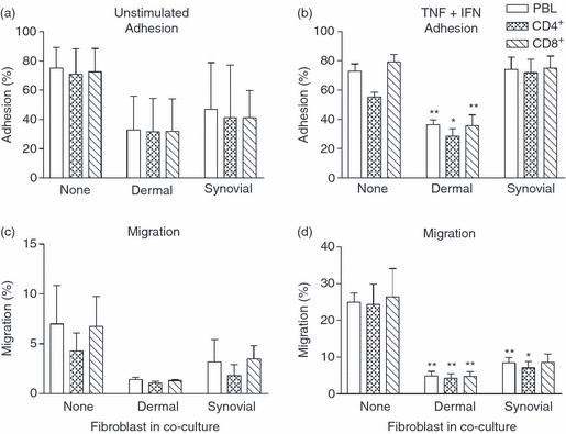
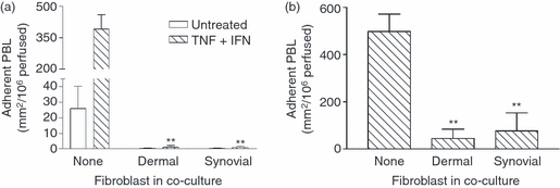
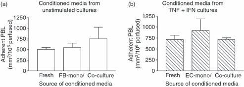
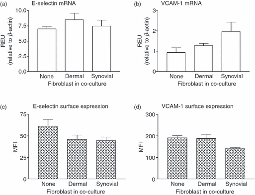
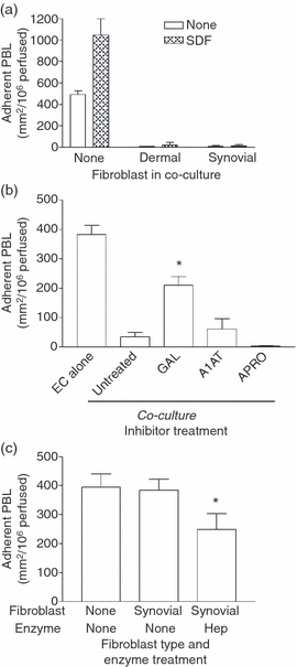
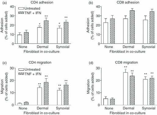
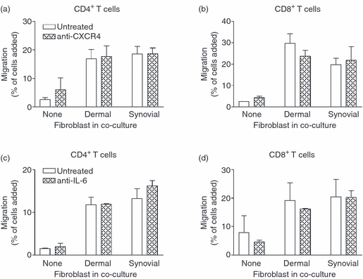
Stromal fibroblasts modify the initial recruitment of leucocytes by endothelial cells (EC), but their effects on subsequent transendothelial migration remain unclear. Here, EC and dermal or synovial fibroblasts were cultured on opposite surfaces of 3-μm pore filters and incorporated in static or flow-based migration assays. Fibroblasts had little effect on tumour necrosis factor-α-induced transendothelial migration of neutrophils, but tended to increase the efficiency of migration away from the endothelium. Surprisingly, similar close contact between EC and fibroblasts strongly reduced lymphocyte migration in static assays, and nearly abolished stable lymphocyte adhesion from flow. Fibroblasts did not alter endothelial surface expression of adhesion molecules or messenger RNA for chemokines. Inhibition of attachment did not occur when EC-fibroblast contact was restricted by using 0.4-μm pore filters, but under these conditions pre-treatment with heparinase partially inhibited adhesion. In the 3-μm pore co-cultures, inhibition of metalloproteinase activity partially recovered lymphocyte adhesion, but addition of CXCL12 (SDF-1α) to the endothelial surface did not. Hence, the ability of EC to present activating chemokines for lymphocytes may have been enzymatically inhibited by direct contact with fibroblasts. To avoid contact, we cultured EC and fibroblasts on separate 3-μm pore filters one above the other. Here, fibroblasts promoted the transendothelial migration of lymphocytes. Fibroblasts generate CXCL12, but blockade of CXCL12 receptor had no effect on lymphocyte migration. While stromal cells can provide signal(s) promoting leucocyte migration away from the sub-endothelial space, direct cell contact (which might occur in damaged tissue) may cause disruption of chemokine signalling, specifically inhibiting lymphocyte rather than neutrophil recruitment.


Figures

Figure 1
Figure 1
Neutrophil adhesion and transmigration through endothelial cells (EC) cultured with fibroblasts under static conditions. Fibroblasts were cultured with EC for 24 hr and left untreated or stimulated with 100 U/ml tumour necrosis factor-α (TNF) for the last 4 hr. (a) Adhesion and (b) transmigration were analysed 2 hr after the addition of neutrophils and expressed as percentages of neutrophils added. Analysis of variance (anova) showed a significant effect of TNF stimulation on neutrophil adhesion and transmigration (P < 0·05), but no effect of fibroblasts. (c) Neutrophil migration through co-cultures established for 4 days before assay. anova showed an effect of fibroblasts with borderline significance, P = 0·06. Data are mean ± SEM from three or four independent experiments.
Figure 2
Figure 2
Adhesion and behaviour of flowing neutrophils on cytokine-treated co-cultures. Endothelial cells cultured alone or with fibroblasts for 24 hr were subsequently stimulated with tumour necrosis factor-α (TNF) for 4 hr before perfusion of a 4-min bolus of neutrophils. (a) Number of neutrophils adherent to co-cultures 2 min after washout. (b) Behaviour of adherent neutrophils at 11 min after washout. (c) Location of migrated neutrophils above or below the filter at 11 min expressed as a percentage of total transmigration. In (c), analysis of variance shows a significant effect of culture conditions on the percentage of migrated neutrophils found above or below the filter, P < 0·01. The percentage of migrated neutrophils below the filter tended to be higher for co-cultures with dermal fibroblasts than for monocultures; P = 0·065 by paired t-test. Data are mean ± SEM from three independent experiments.
Figure 3
Figure 3
Effect of fibroblasts on the adhesion and migration of T-cell subsets under static conditions. Endothelial cells cultured alone or with fibroblasts for 24 hr and were subsequently left untreated (a, c) or stimulated with tumour necrosis factor-α plus interferon-γ (TNF + IFN) (b, d). Adhesion (a, b) and migration (c, d) were assessed at 24 hr and expressed as a percentage of cells added for peripheral blood lymphocytes (PBL), CD4+ or CD8+ T cells. Data are mean ± SEM from three independent experiments. In (b), analysis of variance (anova) shows a significant effect of fibroblasts on T-cell adhesion, P < 0·01. In (c) and (d), anova shows a significant effect of fibroblasts on migration, P < 0·05. * = P < 0·05 and ** = P < 0·01 compared with None by Dunnett test.
Figure 4
Figure 4
Effect of fibroblasts on lymphocyte recruitment from flow. (a) Fibroblasts were cultured alone for 24 hr, and then with endothelial cells (EC) for 24 hr; co-cultures were then left untreated or stimulated with tumour necrosis factor-α plus interferon-γ (TNF + IFN) for 24 hr. (b) EC were cultured alone for 4 days, then with fibroblasts for 24 hr, and then TNF + IFN were added for 24 hr. Data are mean ± SEM from four independent experiments. In (a) and (b), analysis of variance showed a significant effect of fibroblasts on lymphocyte adhesion (P < 0·01). ** = P < 0·01 compared to Untreated or None by Dunnett test.
Figure 5
Figure 5
Effect of conditioned media on lymphocyte recruitment to endothelial cells (EC) from flow. Mono- or co-cultures were formed for 24 hr before treatment with or without cytokines for a further 24 hr, after which the conditioned medium was collected. (a) EC were cultured in fresh medium (Fresh) or in conditioned medium from unstimulated fibroblasts cultured alone (FB-mono) or with EC (Co-culture), with tumour necrosis factor-α plus interferon-γ (TNF + IFN) added for 24 hr. (b) Endothelial cells were cultured for 24 hr in fresh medium with TNF + IFN added (Fresh) or in conditioned medium from EC which had been cultured alone (EC-mono) or with fibroblasts (Co-culture) for 24 hr with TNF + IFN. Data are mean ± SEM from three independent experiments.
Figure 6
Figure 6
Effect of fibroblasts on the expression of E-selectin or vascular cell adhesion molecule 1 (VCAM-1) by endothelial cells (EC). Co-cultures were formed for 24 hr before treatment with tumour necrosis factor-α plus interferon-γ (TNF + IFN) for a further 24 hr. In (a) and (b), endothelial messenger RNA was isolated from co-cultures treated with TNF + IFN and the gene expression of E-selectin (a) or VCAM-1 (b) was assessed by quantitative polymerase chain reaction. Values were expressed as relative expression units (REU) compared with β-actin. In (c) and (d), EC from cytokine-treated co-cultures were labelled using antibodies against (c) E-selectin or (d) VCAM-1 and surface expression was assessed by flow cytometry (expressed as mean fluorescence intensity; MFI). Data are mean ± SEM from three independent experiments.
Figure 7
Figure 7
Effects of modifying chemokine presentation on adhesion of flowing lymphocytes to endothelial cells (EC) cultured alone or with fibroblasts. (a) Co-cultures were formed for 24 hr before treatment with tumour necrosis factor-α plus interferon-γ (TNF + IFN) for 24 hr and then 100 ng/ml CXCL12 was added to EC for 30 min before assay. (b) EC were cultured alone or with synovial fibroblasts on either side of 3·0-μm pore filters for 24 hr and treated with TNF + IFN for 24 hr in the presence or absence of 10 μm galardin (GAL), 100 μg/ml α1-antitrypsin (A1AT) or 50 μg/ml aprotinin (APRO). (c) EC were cultured alone (none) or with synovial fibroblasts on either side of 0·4 um pore filters for 24 hr, treated with TNF + IFN for 24 hr and then 10 mU/ml heparinase (HEP) was added for 30 min. Data are the mean ± SEM from two (a) or three (b, c) independent experiments. * = P < 0·05 compared with synovial None by paired t-test.
Figure 8
Figure 8
Adhesion and migration of lymphocytes through co-cultures in a two-filter static model. Co-cultures were established by inserting an endothelial-coated 24-well filter into a fibroblast-coated 12-well filter. Cells were cultured together for 24 hr and then with or without tumour necrosis factor-α plus interferon-γ (TNF + IFN) for a further 24 hr. T-cell adhesion (a, b) and migration (c, d) were assessed after 10 min and at 24 hr, respectively, using flow cytometry. Data were expressed as a percentage of cells added. In (a) and (c), analysis of variance (anova) shows a significant effect of fibroblasts on CD4+ T-cell adhesion (P < 0·05) and migration (P < 0·01). In (d), anova shows a significant effect of fibroblasts on CD8+ T-cell migration (P < 0·001). Data are the mean ± SEM from six to nine (untreated) and 12 (TNF + IFN) independent experiments. * = P < 0·05 and ** = P < 0·01 compared with endothelial cells cultured alone (none) with matched cytokine treatment by Dunnett test.
Figure 9
Figure 9
Effects of inhibiting actions of CXCL12 or interleukin-6 (IL-6) on migration of T cells through cytokine-treated co-cultures in a two-filter static model. Co-cultures were established by inserting an endothelial-coated 24-well filter into a fibroblast-coated 12-well filter. Cells were cultured together for 24 hr and then with tumour necrosis factor-α plus interferon-γ (TNF + IFN) for a further 24 hr. In (a) and (b), peripheral blood lymphocytes were untreated or treated with the CXCR4 inhibitor AMD3100 for 30 min before the assay. In (c) and (d), neutralizing antibody against IL-6 was added upon establishment of co-culture and was present throughout cytokine-treatment. CD4+ and CD8+ T-cell migration was assessed at 24 hr by flow cytometry and expressed as a percentage of cells added. Data are the mean ± SEM from two (a–b) or three (c–d) independent experiments.

Similar articles

Cited by

References

    1. Ley K, Laudanna C, Cybulsky MI, Nourshargh S. Getting to the site of inflammation: the leukocyte adhesion cascade updated. Nat Rev Immunol. 2007;7:678–89. - PubMed
    1. Luu NT, Rainger GE, Nash GB. Differential ability of exogenous chemotactic agents to disrupt transendothelial migration of flowing neutrophils. J Immunol. 2000;164:5961–9. - PubMed
    1. Tull SP, Yates CM, Maskrey BH, et al. Omega-3 Fatty acids and inflammation: novel interactions reveal a new step in neutrophil recruitment. PLoS Biol. 2009;7:e1000177. - PMC - PubMed
    1. Luscinskas FW, Ding H, Lichtman AH. P-selectin and vascular cell adhesion molecule 1 mediate rolling and arrest, respectively, of CD4+ T lymphocytes on tumour necrosis factor-a-activated vascular endothelium under flow. J Exp Med. 1995;181:1179–86. - PMC - PubMed
    1. Piali L, Weber C, LaRosa G, Mackay CR, Springer TA, Clark-Lewis I, Moser B. The chemokine receptor CXCR3 mediates rapid and shear-resistant adhesion-induction of effector T lymphocytes by the chemokines IP10 and Mig. Eur J Immunol. 1998;28:961–72. - PubMed

Publication types

MeSH terms