. 2010 Dec 6;7(53):1699-707.
doi: 10.1098/rsif.2010.0201. Epub 2010 Jun 2.

Function of blue iridescence in tropical understorey plants

Affiliations

Function of blue iridescence in tropical understorey plants

Katherine R Thomas et al. J R Soc Interface. .

Abstract

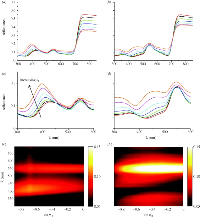
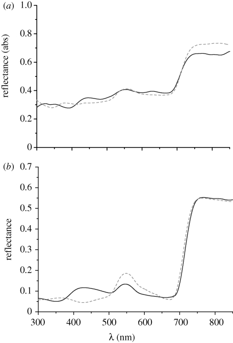
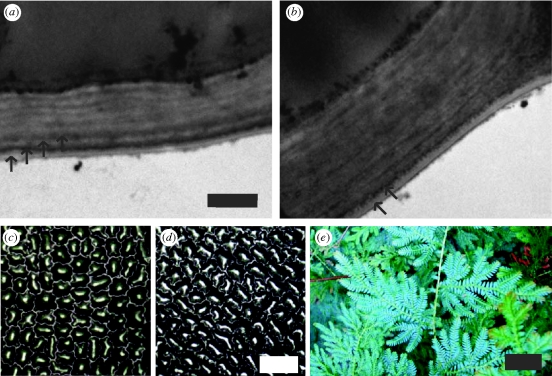
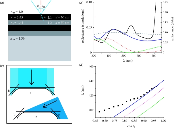
The blue colouration seen in the leaves of Selaginella willdenowii is shown to be iridescent. Transmission electron microscopy studies confirm the presence of a layered lamellar structure of the upper cuticle of iridescent leaves. Modelling of these multi-layer structures suggests that they are responsible for the blue iridescence, confirming the link between the observed lamellae and the recorded optical properties. Comparison of blue and green leaves from the same plant indicates that the loss of the blue iridescence corresponds to a loss of the multi-layer structure. The results reported here do not support the idea that iridescence in plants acts to enhance light capture of photosynthetically important wavelengths. The reflectance of light in the range 600-700 nm is very similar for both iridescent and non-iridescent leaves. However, owing to the occurrence of blue colouration in a wide variety of shade dwelling plants it is probable that this iridescence has some adaptive benefit. Possible adaptive advantages of the blue iridescence in these plants are discussed.


Figures

Figure 1.
Figure 1.
Specular reflection and scattering spectra of (a,c,e) juvenile blue and (b,d,f) mature green S. willdenowii leaves. Intensity of specularly reflected light as a function of light incidence angle for (a) juvenile blue leaves and (b) mature green leaves (red line, θ1 = 16°; black line, θ1 = 22°; green line, θ1 = 28°; blue line, θ1 = 34°; light blue line, θ1 = 40°; purple line, θ1 = 46°; yellow line, θ1 = 50°). (c,d) Enlargements of the reflection peaks in the blue and green spectral ranges from (a,b), respectively. Scattered intensity as a function of detection angle for a fixed light incidence angle of 36° for (e) juvenile blue leaves and (f) mature green leaves. The intensity is colour coded, with red (dark) corresponding to a lower intensity and yellow (bright) a higher intensity.
Figure 2.
Figure 2.
(a) Integrating sphere reflection measurements of blue (solid line) and mature green (dashed line) S. willdenowii leaves. (b) Spectrally resolved specular reflection of juvenile blue (solid line) and mature green (dashed line) leaves at θI = −θD = 16°.
Figure 3.
Figure 3.
TEM micrographs of the outer cell wall and the cuticle from the upper epidermis of (a) a juvenile blue leaf and (b) an older green leaf (scale bar, 500 nm). Arrows indicate the layers observed in the cuticle. Optical micrographs of surface morphology of plant cells for (c) a juvenile blue leaf and (d) a mature green leaf (scale bar, 50 µm). (e) Photograph of juvenile S. willdenowii leaves (scale bar, 30 mm).
Figure 4.
Figure 4.
(a) Multi-layer model showing the thickness and refractive indices of the layers. (b,d) Comparison of experimental data from the juvenile blue leaf and simulated reflectance from an optical multi-layer with d1 = 66 nm and d2 = 50 nm, assuming different combinations of n1 and n2. (b) The dashed line shows simulations for a simplified leaf structure with a single layer of n = 1.45, d = 100 nm on top of a homogeneous optical medium with n = 1.36. The dotted and dashed-dotted lines show simulated reflectance for the simplified leaf covered by an antireflective (AR) coating with n = 1.24 optimized for (dotted) 650 nm and (dashed-dotted) 550 nm, respectively (solid black line, experimental data; solid dark blue line, n1 = 1.45, n2 = 1.44, nout = 1.36; dashed grey line, simplified leaf structure; red dotted line, AR coating 650 nm, n = 1.24, d = 130 nm; green dashed-dotted line, AR coating 550 nm, n = 1.24, d = 110 nm). (c) Contributions from faces a, b and c for light with normal incidence and light with a higher incident angle. (d) Wavelength of the peak in the reflectance between 390 and 450 nm. The solid line shows the theoretical peak shift as calculated using (a). The dotted and dashed lines show the theoretical peak shift for other combinations of n1 and n2. Black points show experimental data from figure 1b. Each data point is an average of three measurements (filled black squares, experimental data; solid dark blue line, four layers n1 = 1.45, n2 = 1.44; purple dotted line, four layers n1 = 1.45, n2 = 1.43; green dashed line, four layers n1 = 1.45, n2 = 1.42).

Similar articles

Cited by

References

    1. Abbott I. A. 1971. On the species of Iridaea (Rhodophyta) from the Pacific coast of North America. Syesis 4, 51–72.
    1. Bazzaz F. A., Pickett S. T. A. 1980. Physiological ecology of tropical succession—a comparative review. Annu. Rev. Ecol. Syst. 11, 287–310. (10.1146/annurev.es.11.110180.001443) - DOI
    1. Blakenship R. E. 2002. Molecular mechanisms of photosynthesis. Oxford, UK: Blackwell Science Ltd.
    1. Charney E., Brackett F. S. 1961. Spectral dependence of scattering from a spherical alga and its implications for the state of organization of the light-accepting pigments. Arch. Biochem. Biophys. 92, 1–12. (10.1016/0003-9861(61)90210-7) - DOI - PubMed
    1. Chazdon R. L., Pearcy R. W., Lee D. W., Fletcher N. 1996. Photosynthetic responses of tropical forest plants to contrasting light environments. In Tropical forest plant ecophysiology (eds Mulkey S. S., Chazdon R. L., Smith A. P.). Boca Raton, FL: Chapman and Hall.

Publication types

LinkOut - more resources