

Biomechanical properties of single chondrocytes and chondrons determined by micromanipulation and finite-element modelling
- PMID: 20519215
- PMCID: PMC2988268
- DOI: 10.1098/rsif.2010.0207
Biomechanical properties of single chondrocytes and chondrons determined by micromanipulation and finite-element modelling
Abstract
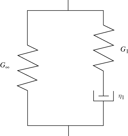
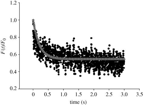
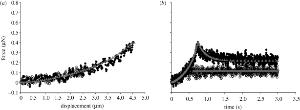
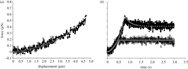
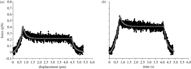
A chondrocyte and its surrounding pericellular matrix (PCM) are defined as a chondron. Single chondrocytes and chondrons isolated from bovine articular cartilage were compressed by micromanipulation between two parallel surfaces in order to investigate their biomechanical properties and to discover the mechanical significance of the PCM. The force imposed on the cells was measured directly during compression to various deformations and then holding. When the nominal strain at the end of compression was 50 per cent, force relaxation showed that the cells were viscoelastic, but this viscoelasticity was generally insignificant when the nominal strain was 30 per cent or lower. The viscoelastic behaviour might be due to the mechanical response of the cell cytoskeleton and/or nucleus at higher deformations. A finite-element analysis was applied to simulate the experimental force-displacement/time data and to obtain mechanical property parameters of the chondrocytes and chondrons. Because of the large strains in the cells, a nonlinear elastic model was used for simulations of compression to 30 per cent nominal strain and a nonlinear viscoelastic model for 50 per cent. The elastic model yielded a Young's modulus of 14 ± 1 kPa (mean ± s.e.) for chondrocytes and 19 ± 2 kPa for chondrons, respectively. The viscoelastic model generated an instantaneous elastic modulus of 21 ± 3 and 27 ± 4 kPa, a long-term modulus of 9.3 ± 0.8 and 12 ± 1 kPa and an apparent viscosity of 2.8 ± 0.5 and 3.4 ± 0.6 kPa s for chondrocytes and chondrons, respectively. It was concluded that chondrons were generally stiffer and showed less viscoelastic behaviour than chondrocytes, and that the PCM significantly influenced the mechanical properties of the cells.
Figures

Similar articles
-
Strain-dependent viscoelastic behaviour and rupture force of single chondrocytes and chondrons under compression.Biotechnol Lett. 2009 Jun;31(6):803-9. doi: 10.1007/s10529-009-9939-y. Epub 2009 Feb 11. Biotechnol Lett. 2009. PMID: 19205892
-
Zonal uniformity in mechanical properties of the chondrocyte pericellular matrix: micropipette aspiration of canine chondrons isolated by cartilage homogenization.Ann Biomed Eng. 2005 Oct;33(10):1312-8. doi: 10.1007/s10439-005-4479-7. Ann Biomed Eng. 2005. PMID: 16240080
-
An axisymmetric boundary element model for determination of articular cartilage pericellular matrix properties in situ via inverse analysis of chondron deformation.J Biomech Eng. 2010 Mar;132(3):031011. doi: 10.1115/1.4000938. J Biomech Eng. 2010. PMID: 20459199 Free PMC article.
-
The deformation behavior and mechanical properties of chondrocytes in articular cartilage.Osteoarthritis Cartilage. 1999 Jan;7(1):59-70. doi: 10.1053/joca.1998.0162. Osteoarthritis Cartilage. 1999. PMID: 10367015 Review.
-
The deformation behavior and viscoelastic properties of chondrocytes in articular cartilage.Biorheology. 2000;37(1-2):27-44. Biorheology. 2000. PMID: 10912176 Review.
Cited by
-
Ex Vivo Systems to Study Chondrogenic Differentiation and Cartilage Integration.J Funct Morphol Kinesiol. 2021 Jan 5;6(1):6. doi: 10.3390/jfmk6010006. J Funct Morphol Kinesiol. 2021. PMID: 33466400 Free PMC article. Review.
-
Combination of chondrocytes and chondrons improves extracellular matrix production to promote the repairs of defective knee cartilage in rabbits.J Orthop Translat. 2021 Feb 23;28:47-54. doi: 10.1016/j.jot.2021.01.004. eCollection 2021 May. J Orthop Translat. 2021. PMID: 33717981 Free PMC article.
-
Regional variations in growth plate chondrocyte deformation as predicted by three-dimensional multi-scale simulations.PLoS One. 2015 Apr 17;10(4):e0124862. doi: 10.1371/journal.pone.0124862. eCollection 2015. PLoS One. 2015. PMID: 25885547 Free PMC article.
-
Microscale mapping of extracellular matrix elasticity of mouse joint cartilage: an approach to extracting bulk elasticity of soft matter with surface roughness.Soft Matter. 2018 Apr 18;14(15):2879-2892. doi: 10.1039/c7sm02045g. Soft Matter. 2018. PMID: 29582024 Free PMC article.
-
Tribological and Rheological Properties of Poly(vinyl alcohol)-Gellan Gum Composite Hydrogels.Polymers (Basel). 2022 Sep 14;14(18):3830. doi: 10.3390/polym14183830. Polymers (Basel). 2022. PMID: 36145975 Free PMC article.
References
-
- ABAQUS 2005. Analysis user's manual, v. 6.5. Pawtucket, RI: Hibbit, Karlsson and Sorensen Inc.
-
- Andrei D. C., Briscoe B. J., Luckham P. F., Williams D. R. 1996. The deformation of microscopic gel particles. J. Chim. Phys. Phys. Chim. Biol. 93, 960–976.
Publication types
MeSH terms
LinkOut - more resources
Full Text Sources
Other Literature Sources
