Review
. 2010 Sep 1;588(Pt 17):3187-200.
doi: 10.1113/jphysiol.2010.191973. Epub 2010 Jun 2.

Going native: voltage-gated potassium channels controlling neuronal excitability

Affiliations
Review

Going native: voltage-gated potassium channels controlling neuronal excitability

Jamie Johnston et al. J Physiol. .

Abstract

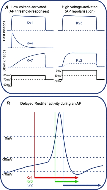
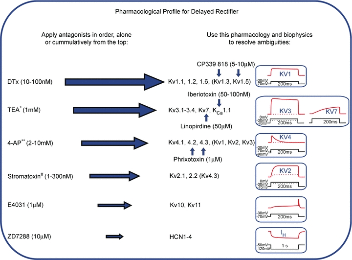
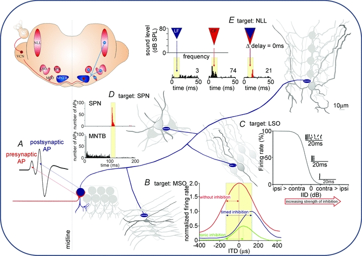
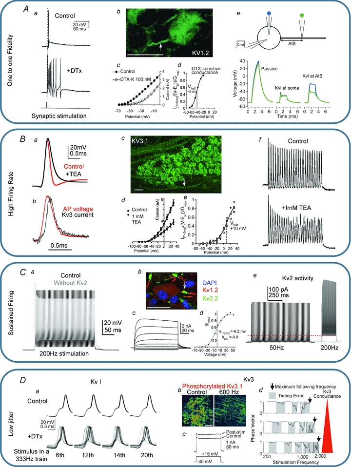
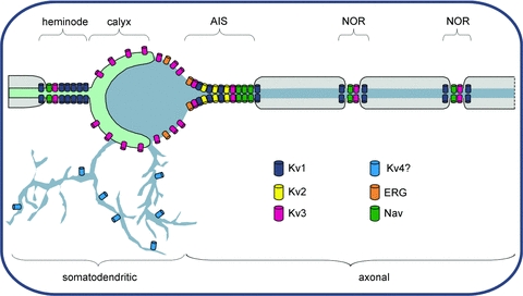
In this review we take a physiological perspective on the role of voltage-gated potassium channels in an identified neuron in the auditory brainstem. The large number of KCN genes for potassium channel subunits and the heterogeneity of the subunit combination into K(+) channels make identification of native conductances especially difficult. We provide a general pharmacological and biophysical profile to help identify the common voltage-gated K(+) channel families in a neuron. Then we consider the physiological role of each of these conductances from the perspective of the principal neuron in the medial nucleus of the trapezoid body (MNTB). The MNTB is an inverting relay, converting excitation generated by sound from one cochlea into inhibition of brainstem nuclei on the opposite side of the brain; this information is crucial for binaural comparisons and sound localization. The important features of MNTB action potential (AP) firing are inferred from its inhibitory projections to four key target nuclei involved in sound localization (which is the foundation of auditory scene analysis in higher brain centres). These are: the medial superior olive (MSO), the lateral superior olive (LSO), the superior paraolivary nucleus (SPN) and the nuclei of the lateral lemniscus (NLL). The Kv families represented in the MNTB each have a distinct role: Kv1 raises AP firing threshold; Kv2 influences AP repolarization and hyperpolarizes the inter-AP membrane potential during high frequency firing; and Kv3 accelerates AP repolarization. These actions are considered in terms of fidelity of transmission, AP duration, firing rates and temporal jitter. An emerging theme is activity-dependent phosphorylation of Kv channel activity and suggests that intracellular signalling has a dynamic role in refining neuronal excitability and homeostasis.


Figures

Figure 1
Figure 1. Functional classification of voltage-gated potassium currents
A, voltage clamp permits the biophysical properties of the native channel types to be distinguished in terms of voltage-dependent activation kinetics. Generalization of these properties is expressed here as a matrix of low-voltage-activated (LVA) channels such as Kv1, Kv4 and Kv7, and high-voltage-activated channels (HVA) Kv3 and Kv2. Kv4 channels (mediating A-type currents) are inactivated at rest and so require prior hyperpolarization before they will activate. The spectrum of their channel kinetics further divides these groups into those that activate rapidly (Kv1, Kv4 and Kv3) and more slowly (Kv7 and Kv2). B, under conventional voltage recording (current clamp) the Kv channel kinetics mean that each channel activates over a characteristic part of the overall AP waveform as represented by the coloured bars/arrows: LVA channels open on depolarization from resting potentials (−70 mV) to around −40 to −30 mV, so influencing the threshold for AP firing. HVA channels require further depolarization, approaching 0 mV, which is only achieved during APs, so these channels contribute to repolarization. Kv1 channels would turn on with small depolarizations, while Kv3 would be delayed until further depolarization. Kv2 would be even later due to its slow kinetics, but its activity extended to longer time points as it is slow to turn off. Although this diagram is a vast oversimplification, it gives a framework in which to identify native potassium currents and their different roles in determining neuronal excitability. Note that little emphasis is placed on inactivation, since this is so dependent on the precise subunit composition of the channel, but it is nonetheless important; Kv4 channels invariably inactivate, with a time course which is highly dependent on subunit and accessory proteins. Inactivation (for reviews see Robertson, 1997; Aldrich, 2001) plays an important role in reducing the Kv contribution during sustained activation, for instance increasing AP duration during repetitive firing.
Figure 2
Figure 2. Pharmacological profile for native delayed rectifier Kv families
Since native channels are often heteromers, precise biophysical and pharmacological matching to recombinant homomeric channels is difficult. Measurement of ion channel current defines the functional plasma membrane channel pool; this is a crucial advantage over biochemical approaches (Western blot or PCR). This pharmacological profile method should be applied with caution. The antagonists (or gating modifier no.) listed on the left are applied cumulatively in order to inhibit the conductances indicated in the middle. Resolution of ambiguous pharmacology is achieved by secondary experiments using the additional pharmacology indicated by the vertical blue arrows. Additional constraints and confirmation of Kv identity is obtained from the time course and current–voltage relations of the current measured under voltage clamp, as summarized on the right. *Low concentrations of TEA will also block Kv1.1 homomeric channels. ** 4-aminopyridine will block many Kv1 and Kv3 subunits at micromolar concentrations and most Kvs at millimolar concentrations; If Kv4 (A-current) is present it may be effectively removed by induction of inactivation, without need of pharmacological agents. #Stromatoxin is a gating modifer, shifting the activation voltage to more positive potentials, rather than an antagonist.
Figure 3
Figure 3. Contribution of MNTB-inhibition to auditory brainstem processing
Inset, a sketch of a coronal brain section at the level of the superior olivary complex. Sound-evoked activity from the contralateral ear arrives as AP trains in the ventral cochlear nucleus (VCN) which sends excitatory projections to the MNTB (red lines, +). MNTB neurons give inhibitory projections (blue lines, −) to the medial superior olive (MSO), the lateral superior olive (LSO), the superior paraolivary nucleus (SPN) and the nuclei of the lateral lemniscus (NLL). All four target nuclei receive direct excitatory inputs from the VCN (not shown). A, excitation to each MNTB neuron is mediated via a calyx of Held synapse (red line) whose activity can be detected in extracellular recordings as a presynaptic AP preceding the postsynaptic AP (modified from Kopp-Scheinpflug et al. 2008b with permission from Elsevier). B, MSO cells receive glycinergic IPSPs directly onto their somata. Without inhibition, the peak of the ITD function lies within the physiologically relevant range of ITDs (yellow shaded area) and ITDs are encoded by a topographic map. Well-timed inhibition reduces the firing rate but also shifts the peak ITD function out of the physiological range. In this scenario the slope of the ITD function can be used to encode ITDs via a rate code. Tonic (non-phase-locked) inhibition causes a reduction in firing rate but without the respective shift of the ITD function (figure modified from Pecka et al. 2008 with permission from the Society for Neuroscience). C, LSO cells respond to ipsilateral sound stimulation with increased firing rates. Increasing the strength of the inhibitory input leads to successive reductions in firing rate. Dot raster plots resemble the activity at about 20, 50 and 80% reduction of firing rate. The inhibition needs to be rather strong to suppress the onset component (figure modified from Park et al. 1997, with permission from the American Physiological Society). D, SPN cells receive a sustained inhibitory input which effectively suppresses AP firing during sound stimulation. The termination of a sound is then highlighted by an AP offset response (unpublished data; see also Kadner et al. 2006). E, NLL neurons receive low-frequency inhibition (blue area and histogram below), in addition to a higher-frequency excitatory CF-stimulation (red area and histogram below). When both stimuli are presented at 0 ms delay, the excitatory CF response is suppressed (overlaying blue and red areas) and a post-inhibitory rebound is observed (red part of the histogram; figure modified from Peterson et al. 2009 with permission from the American Physiological Society).
Figure 4
Figure 4. K+ channel physiology of the MNTB
A, Kv1 channels ensure one-to-one fidelity. Aa, application of DTx results in multiple action potentials on the tail of the EPSP generated in response to each presynaptic stimulus (Brew & Forsythe, 1995). Ab, Kv1 immunohistochemistry shows that Kv1 channels are located in the axon initial segment (Dodson et al. 2002); scale bar is 20 μm. Ac and d, DTx blocks a LVA K+ current with a V0.5 of −43 mV (Brew & Forsythe, 1995; Dodson et al. 2002 with permission, SfN.). Ae, a simple NEURON model (Hines & Carnevale, 2001) of the MNTB with a 1.8 nA current injection at the soma. The voltage at the soma and at the end of the AIS is plotted in blue and green, respectively, and shown assuming passive properties (left) Kv1 channels located at the soma (middle) and with Kv1 channels in the AIS (right). Note that with Kv1 channels in the AIS the voltage in the soma differs from the AIS, which explains the apparent lowering of action potential threshold seen with DTx application (Dodson et al. 2002). B, Kv3 channels ensure high firing rates. Ba, blocking Kv3 channels with TEA slows action potential repolarization. Bb, Kv3 current activates with the action potential peaking at the start of repolarization (reproduced from Klug & Trussell, 2006 with permission from the American Physiological Society). Bc, Kv3 immunohistochemistry shows these channels in the MNTB cell body and in the axon (arrow indicates nodes of Ranvier); scale bar is 40 μm. Bd and e, 1 mm TEA blocks a high voltage-activated K+ current with a V0.5 of ∼10 mV. Bf, the Kv3 mediated rapid repolarization is essential for high firing rates (Wang et al. 1998). C, Kv2 channels enable sustained firing. Ca, in a single compartment NEURON model, removing Kv2 results in a depolarized inter-spike potential and reduced availability of Nav channels causing shorter APs. Cb, Kv2 channels are located in the AIS along with Kv1 channels; scale bar is 20 μm. Cc and d, Kv2 channels mediate a slow activating high voltage-activated K+ current. Ce, Kv2 current is activated in a frequency-dependent manner and remains active during the inter-spike potential (Johnston et al. 2008a). D, decreased jitter with Kv1 channels and regulation of Kv3 channels. Da, Kv1 channels decrease the membrane time constant and improve the timing of MNTB neurons during repetitive stimulation (reproduced from Gittelman & Tempel, 2006 with permission from the American Phsyiological Society). Db, Kv3 phosphorylation state is regulated by activity; scale bar is 100 μm. Dc, dephosphorylation occurs in loud auditory environments or with high frequency stimulation and increases Kv3 current magnitude. Dd, large Kv3 magnitudes prolong firing but introduce jitter (Song et al. 2005 reproduced with permission, Nature Neuroscience); under quiet conditions Kv3 is phosphorylated and timing errors are minimized.
Figure 5
Figure 5. Subcellular localization of voltage-gated K+ channels
Somatic Kv channels include Kv3 and ERG, but the major part of the conductance arises from channels in the axon initial segment (AIS) which are dominated by Kv1, Kv2 and Kv3 channels (as well as voltage-gated sodium channels, Nav). The location of Kv4 on dendrites is implied from their absence in MNTB somatic membrane and evidence from other cell types. Nodes of Ranvier (NOR) contain Nav and Kv3, with Kv1 channels in the juxtaparanodal region, under the myelin sheath (Wang et al. 1993). The last NOR of the axon known as the heminode is particularly large for the Calyx (Leão et al. 2005) and contains Kv1 and Kv3 channels, with the majority of Kv3 being on the non-release face of the synaptic terminal.
None

Similar articles

Cited by

References

    1. Aldrich RW. Fifty years of inactivation. Nature. 2001;411:643–644. - PubMed
    1. Armstrong CM, Binstock L. Anomalous rectification in the squid giant axon injected with tetraethylammonium chloride. J Gen Physiol. 1965;48:859–872. - PMC - PubMed
    1. Banks MI, Smith PH. Intracellular recordings from neurobiotin-labelled cells in brain slices of the rat medial nucleus of the trapezoid body. J Neurosci. 1992;12:2819–2837. - PMC - PubMed
    1. Barnes-Davies M, Forsythe ID. Pre- and postsynaptic glutamate receptors at a giant excitatory synapse in rat auditory brainstem slices. J Physiol. 1995;488:387–406. - PMC - PubMed
    1. Brew HM, Forsythe ID. Two voltage-dependent K+ conductances with complementary functions in postsynaptic integration at a central auditory synapse. J Neurosci. 1995;15:8011–8022. - PMC - PubMed

Publication types

MeSH terms

Substances

LinkOut - more resources