. 2010 Jul 15;588(Pt 14):2621-31.
doi: 10.1113/jphysiol.2010.188979. Epub 2010 Jun 2.

Pre- and postsynaptic modulation of monosynaptic reflex by GABAA receptors on turtle spinal cord

Affiliations

Pre- and postsynaptic modulation of monosynaptic reflex by GABAA receptors on turtle spinal cord

Wendy Bautista et al. J Physiol. .

Abstract

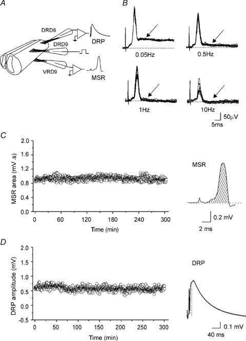
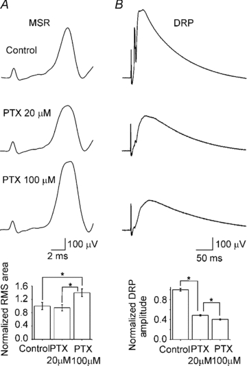
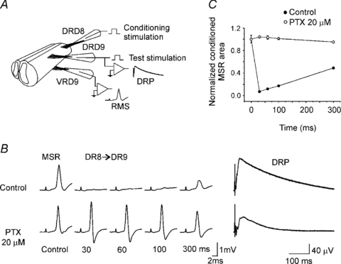
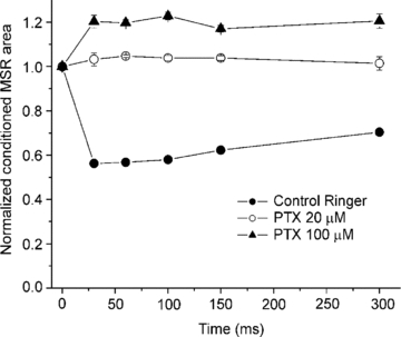
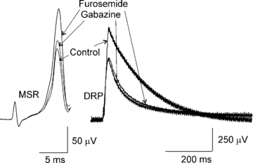
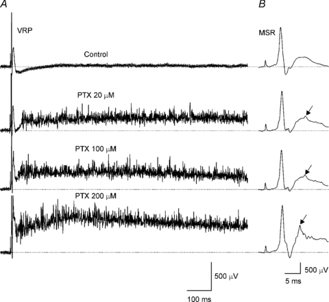
There is growing evidence that activation of high affinity extrasynaptic GABA(A) receptors in the brain, cerebellum and spinal cord substantia gelatinosa results in a tonic inhibition controlling postsynaptic excitability. The aim of the present study was to determine if GABA(A) receptors mediating tonic inhibition participate in the modulation of monosynaptic reflex (MSR) in the vertebrate spinal cord. Using an in vitro turtle lumbar spinal cord preparation, we show that conditioning stimulation of a dorsal root depressed the test monosynaptic reflex (MSR) at long condition-test intervals. This long duration inhibition is similar to the one seen in mammalian spinal cord and it is dependent on GABA(A) as it was completely blocked by 20 microm picrotoxin (PTX) or bicuculline (BIC) or 1 microm gabazine, simultaneously depressing the dorsal root potential (DRP) without MSR facilitation. Interestingly 100 microm picrotoxin or BIC potentiated the MSR, depressed the DRP, and produced a long lasting motoneurone after-discharge. Furosemide, a selective antagonist of extrasynaptic GABA(A) receptors, affects receptor subtypes with alpha(4/6) subunits, and in a similar way to higher concentrations of PTX or BIC, also potentiated the MSR but did not affect the DRP, suggesting the presence of alpha(4/6) GABA(A) receptors at motoneurones. Our results suggest that (1) the turtle spinal cord has a GABA(A) mediated long duration inhibition similar to presynaptic inhibition observed in mammals, (2) GABA(A) receptors located at the motoneurones and primary afferents might produce tonic inhibition of monosynaptic reflex, and (3) GABA(A) receptors modulate motoneurone excitability reducing the probability of spurious and inappropriate activation.


Figures

Figure 1
Figure 1. Simultaneous recordings of the monosynaptic reflex (MSR) and the dorsal root potential (DRP) in the turtle spinal cord
A, scheme of two spinal cord segments in continuity with the dorsal and ventral roots attached to suction electrodes for stimulation and recording of the DRP and VRP. B, VRP evoked at four different frequency stimulations (2T) applied to the ipsilateral dorsal root. The earliest component of VRP is the MSR because it followed one to one frequency stimulation at 10 Hz without jittering and failure. The slower VRP component failed to follow one to one at 1 Hz of dorsal root stimulation. Striped area under the MSR curve (C) and the DRP amplitude (D) recorded simultaneously every 30 s plotted versus time. Traces, average of the MSR and DRP recordings of 10 sweeps.
Figure 2
Figure 2. Action of picrotoxin on the MSR and DRP
A and B, top to bottom, representative MSR and DRP evoked by dorsal root stimulation (2T) and simultaneously recorded in control Ringer solution and in the presence of picrotoxin (PTX) at 20 and 100 μm. Bar graphs, normalized MSR area and DRP amplitude in control Ringer solution and in the presence of PTX. Vertical lines indicate s.e.m. Asterisks indicate statistical difference with respect to the control for the MSR and with respect to the control and between PTX at 20 and 100 μm for the DRP (n = 11; P < 0.05).
Figure 3
Figure 3. Long duration inhibition of the MSR
A, scheme showing the protocol to induce long duration inhibition of the MSR. MSR recorded from D9 ventral root (VRD9) was evoked by stimulation of the ipsilateral dorsal root (DRD9). Conditioning stimulation was applied through D8 dorsal root (DRD8). B, MSR control and conditioned (DR8→DR9) recorded in control Ringer solution (top traces) and in presence of picrotoxin (20 μm; lower traces) at interstimulus intervals indicated bellow. To the right, DRP recorded from DRD9 in control Ringer solution and in the presence of picrotoxin. C, plot of normalized MSR area versus interstimulus interval in control Ringer solution (filled circles) and in presence of picrotoxin (open circles). Vertical bars indicate s.e.m.
Figure 4
Figure 4. Faciliation of the conditioned MSR by higher concentrations of picrotoxin
Plot shows the normalized area of MSR evoked by stimulation of the DRD9 conditioned by stimulation of an adjacent dorsal root (DRD8) at interstimulus intervals of 30, 60, 100, 150 and 300 ms in control Ringer solution (filled circles) and in the presence of picrotoxin at 20 (open circles) and 100 μm (filled triangles). Vertical bars indicate the s.e.m.
Figure 5
Figure 5. Furosemide effect on the MSR and DRP
The MSR and DRP recorded in control Ringer solution, in the presence of gabazine 1 μm, and plus furosemide 200 μm.
Figure 6
Figure 6. Motoneurone after-discharge in the presence of picrotoxin
A, VRP evoked by ipsilateral dorsal root stimulation (2T) recorded, from top to bottom, in control Ringer and in the presence of picrotoxin (PTX) at 20, 100 and 200 μm. B, the MSR from A at a different time and voltage scale. Arrows indicate polysynaptic reflex.

References

    1. Alvarez FJ, Taylor-Blake B, Fyffe RE, De Blas AL, Light AR. Distribution of immunoreactivity for the β2 and β3 subunits of the GABAA receptor in the mammalian spinal cord. J Comp Neurol. 1996;365:392–412. - PubMed
    1. Alaburda A, Russo R, MacAulay N, Hounsgaard J. Periodic high-conductance states in spinal neurons during scratch-like network activity in adult turtles. J Neurosci. 2005;27:6316–6321. - PMC - PubMed
    1. Ataka T, Gu JG. Relationship between tonic inhibitory currents and phasic inhibitory activity in the spinal cord lamina II region of adult mice. Mol Pain. 2006;27:2–36. - PMC - PubMed
    1. Bohlhalter S, Weinmann O, Mohler H, Fritschy JM. Laminar compartmentalization of GABAA-receptor subtypes in the spinal cord: an immunohistochemical study. J Neurosci. 1996;16:283–297. - PMC - PubMed
    1. Chadderton P, Margrie TW, Häusser M. Integration of quanta in cerebellar granule cells during sensory processing. Nature. 2004;428:856–860. - PubMed

Publication types

MeSH terms

LinkOut - more resources