

Review
doi: 10.1101/cshperspect.a000398.
Epub 2010 Jun 2.
Biofilms
Affiliations
- PMID: 20519345
- PMCID: PMC2890205
- DOI: 10.1101/cshperspect.a000398
Item in Clipboard
Review
Biofilms
Cold Spring Harb Perspect Biol.
2010 Jul.
Abstract
The ability to form biofilms is a universal attribute of bacteria. Biofilms are multicellular communities held together by a self-produced extracellular matrix. The mechanisms that different bacteria employ to form biofilms vary, frequently depending on environmental conditions and specific strain attributes. In this review, we emphasize four well-studied model systems to give an overview of how several organisms form biofilms: Escherichia coli, Pseudomonas aeruginosa, Bacillus subtilis, and Staphylococcus aureus. Using these bacteria as examples, we discuss the key features of biofilms as well as mechanisms by which extracellular signals trigger biofilm formation.
Figures
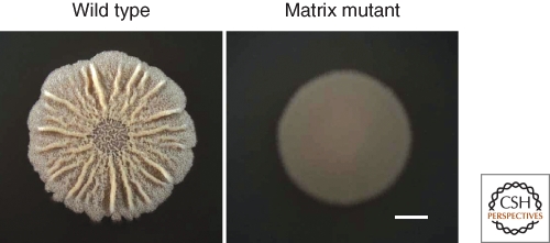
Colony morphology of B. subtilis strain 3610 wild type and matrix mutant (eps). Top view of cells after 3 d of growth on 1.5% agar MSgg medium. Bar is 5 mm.
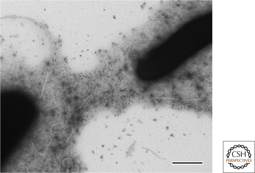
Electron micrograph of B. subtilis strain 3610 immunogold labeled with anti-TasA antibody (black dots). Bar is 0.5 µm. Image courtesy of Diego Romero.
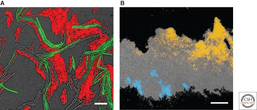
Heterogeneity in B. subtilis biofilms. (A) Top view of cells at the onset of colony development. Overlay of fluorescence images with DIC (gray), motile (red), and matrix-producing (green) cells. Bar 5 µm. (B) Thin-sectioned three-day-old biofilm. Agar is at the bottom and the center of the colony is on the right. Overlay of fluorescence images with DIC (gray), motile (blue), and sporulating (orange) cells. Bar 50 µm.
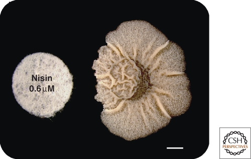
Effect of the antimicrobial nisin on B. subtilis biofilm morphology. Cells closer to the disk containing nisin are more wrinkled due to the presence of more matrix-producing cells. Bar is 3 mm. (Reprinted, with permission, from Lopez et al 2009b [© Wiley].)
References
-
- Allesen-Holm M, Barken KB, Yang L, Klausen M, Webb JS, Kjelleberg S, Molin S, Givskov M, Tolker-Nielsen T 2006. A characterization of DNA release in Pseudomonas aeruginosa cultures and biofilms. Mol Microbiol 59:1114–1128 - PubMed
-
- An D, Parsek MR 2007. The promise and peril of transcriptional profiling in biofilm communities. Curr Opin Microbiol 10:292–296 - PubMed
-
- Anderson GG, O’Toole GA 2008. Innate and induced resistance mechanisms of bacterial biofilms. in Bacterial Biofilms (ed. Romeo T.), pp. 85–105 Springer, Heidelberg - PubMed
Publication types
MeSH terms
LinkOut - more resources
Full Text Sources
Other Literature Sources