. 2010 Aug;151(8):3836-46.
doi: 10.1210/en.2010-0174. Epub 2010 Jun 2.

Neurokinin B acts via the neurokinin-3 receptor in the retrochiasmatic area to stimulate luteinizing hormone secretion in sheep

Affiliations

Neurokinin B acts via the neurokinin-3 receptor in the retrochiasmatic area to stimulate luteinizing hormone secretion in sheep

Heather J Billings et al. Endocrinology. 2010 Aug.

Abstract

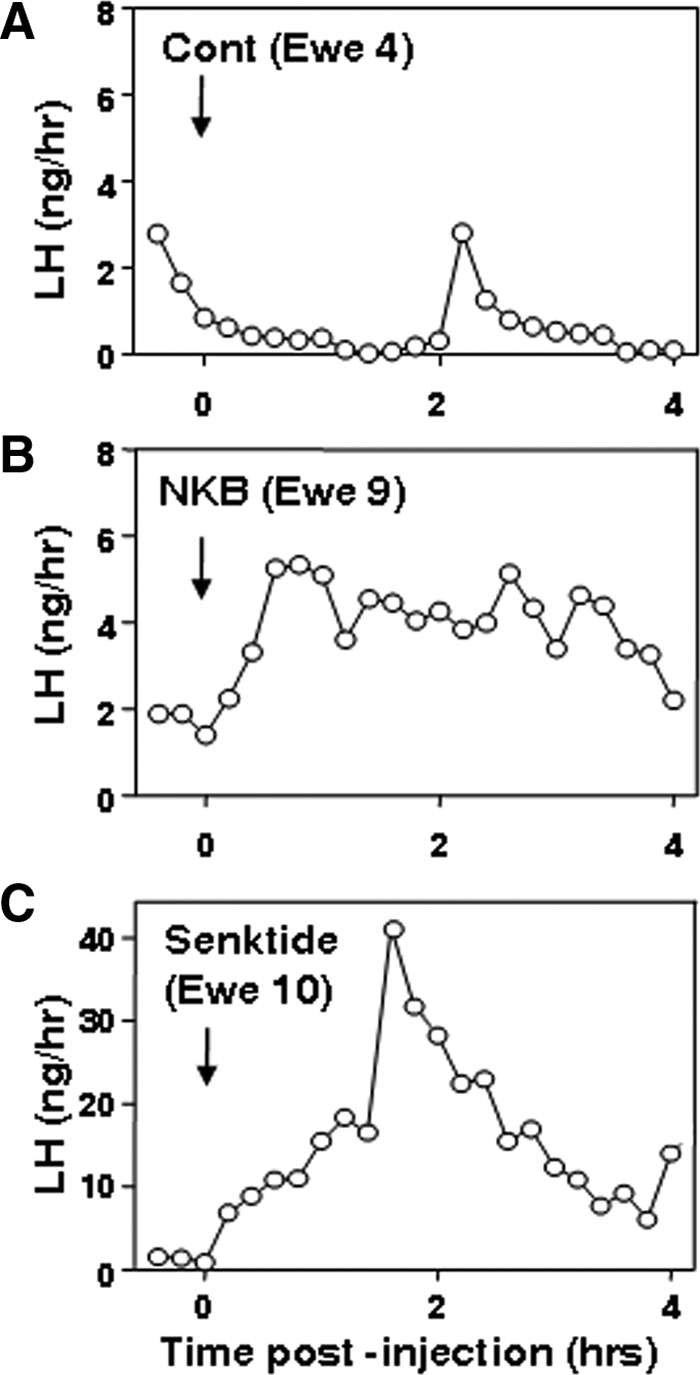
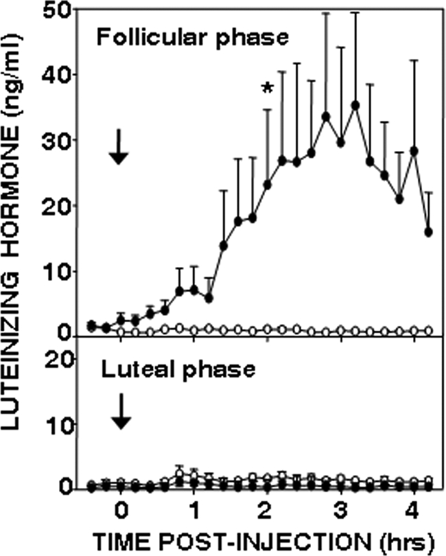
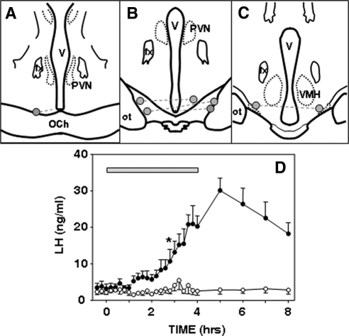
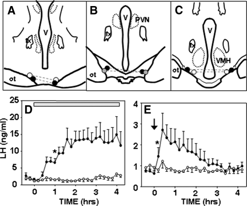
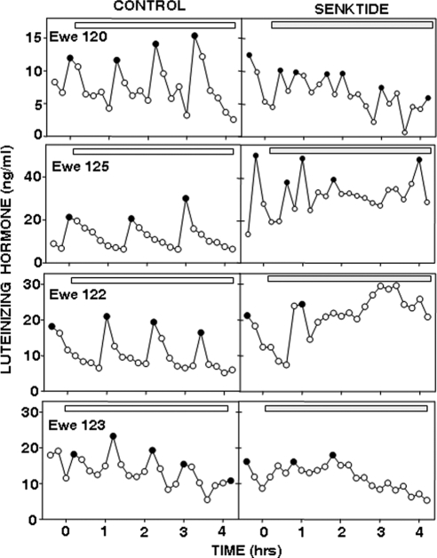
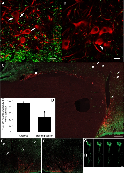
Recent data have demonstrated that mutations in the receptor for neurokinin B (NKB), the NK-3 receptor (NK3R), produce hypogonadotropic hypogonadism in humans. These data, together with reports that NKB expression increases after ovariectomy and in postmenopausal women, have led to the hypothesis that this tachykinin is an important stimulator of GnRH secretion. However, the NK3R agonist, senktide, inhibited LH secretion in rats and mice. In this study, we report that senktide stimulates LH secretion in ewes. A dramatic increase in LH concentrations to levels close to those observed during the preovulatory LH surge was observed after injection of 1 nmol senktide into the third ventricle during the follicular, but not in the luteal, phase. Similar increases in LH secretion occurred after insertion of microimplants containing this agonist into the retrochiasmatic area (RCh) in anestrous or follicular phase ewes. A low-dose microinjection (3 pmol) of senktide into the RCh produced a smaller but significant increase in LH concentrations in anestrous ewes. Moreover, NK3R immunoreactivity was clearly evident in the RCh, although it was not found in A15 dopaminergic cell bodies in this region. These data provide evidence that NKB stimulates LH (and presumably GnRH) secretion in ewes and point to the RCh as one important site of action. Based on these data, and the effects of NK3R mutations in humans, we hypothesize that NKB plays an important stimulatory role in the control of GnRH and LH secretion in nonrodent species.


Figures

Figure 1
Figure 1
LH concentrations after a single icv injection of vehicle (Cont; A), NKB (B), or the NK3R agonist, senktide (C) in individual ovary-intact ewes during the breeding season. Note difference in y-axis in C.
Figure 2
Figure 2
Effect of vehicle (open circles) and senktide (closed circles) injection into the third ventricle in follicular (top panel) and luteal phase (bottom panel) ewes (n = 4). Mean ± sem are shown. There was a significant treatment effect (P < 0.05) and a time × treatment interaction (P < 0.001) in the follicular phase, but not in the luteal phase, based on two-way ANOVA with repeated measures. An asterisk indicates the first time at which there was a statistically significant difference between LH concentrations after senktide and control treatments based on the Tukey test for multiple comparisons. All subsequent comparisons were significant except for the very last time point. There was no effect of senktide on LH pulse frequency (Wilcoxon-Mann-Whitney test) or amplitude (paired t test) in the luteal phase.
Figure 3
Figure 3
Effects of local administration of senktide into the RCh of follicular phase ewes. A–C, Positions of microimplants in schematic coronal sections through the RCh. Bilateral microimplants in each ewe are connected by dashed lines. Note that in one ewe, the microimplant on one side was at the edge of the optic chiasm (OCh), whereas the other side was slightly posterior to it. In the fifth animal, microimplant tracts extended beyond the base of the brain (data not shown). D, Mean LH (±sem, n = 4) concentrations before, during, and after insertion of empty (open circles) and senktide-containing (solid circles) microimplants (horizontal bar) into the RCh. Two-way ANOVA with repeated measures indicated there was a significant treatment effect (P < 0.05) and a time × treatment interaction (P < 0.001). Asterisk indicates the first time at which there was a statistically significant difference between LH concentrations after senktide and control treatments, based on the Tukey test for multiple comparisons. All subsequent comparisons were significant. fx, Fornix; ot, optic tract; PVN, paraventricular nucleus; V, third ventricle; VMH, ventromedial hypothalamus.
Figure 4
Figure 4
Effect of local administration of senktide to the RCh of ovary-intact anestrous ewes. A–C, Positions of microimplants and microinjections in schematic coronal sections through the RCh, with bilateral sites in the same ewe connected by dashed lines. Solid circles denote ewes with a robust response to senktide microinjection (peak LH > 4 ng/ml); shaded circles denote ewe with modest response (peak LH < 2 ng/ml); and open circles denote ewes with no response to senktide microinjection. All six ewes responded to the microimplants. D, Mean (±sem, n = 6) LH concentrations before and after insertion of either empty (open circles) or senktide-containing (closed circles) microimplants (horizontal bar). There was a significant treatment effect (P < 0.01) and a time × treatment interaction (P < 0.001) (two-way ANOVA with repeated measures). E, Mean (±sem, n = 6) LH concentrations before and after bilateral microinjection (arrow) of vehicle (open circles) or 3 pmol senktide (closed circles) into the RCh. There was a significant (P < 0.05) time × treatment interaction (two-way ANOVA with repeated measures). Asterisks in D and E depict the first time at which there was a statistically significant difference between LH concentrations after senktide and control treatments, based on the Tukey test for multiple comparisons. All subsequent comparisons were significantly different in D, but only the next five time points were different after the microinjection (E).
Figure 5
Figure 5
LH pulse patterns in four OVX ewes during anestrus before and after insertion of empty (left panels) and senktide-containing (right panels) microimplants (horizontal bars) into the RCh. Solid circles depict the peak of LH pulses. Note differences in y-axes. Microimplant sites are indicated in Fig. 4, A–C. Ewe 120, shaded circles (B); ewe 122, open circles (C); ewe 123, open circles (A); and ewe 125, solid circles (C).
Figure 6
Figure 6
NK3R and tyrosine hydroxylase dual immunofluorescence labeling in the ovine hypothalamus. A and B, Confocal images of coronal sections through the RCh from OVX+E2 anestrous (A) and breeding season (B) ewes showing NK3R-ir fibers (green) and tyrosine hydroxylase-ir cell bodies (red). Arrows indicate some NK3R-containing varicosities in close contact to A15 cells. Images captured using ×63 objective, ×0.7 optical zoom, and 1.5-μm z-section. Scale bar, 20 μm. C, Montage of confocal images of a parasagittal section illustrating a continuum of NK3R-ir fibers and tyrosine hydroxylase-positive cells along the border of the optic chiasm. Arrows indicate NK3R-positive cell bodies. Images captured using ×10 objective and 15-μm z-thickness. Scale bar, 500 μm. D, Seasonal comparison of the percentage of A15 DA neurons receiving NK3R-containing close contacts. *, P < 0.05. E–H, Confocal images of NK3R-ir neurons (arrows in E) at low (×10 objective and 15-μm z-thickness; E and F) and high (×63 objective and 1-μm z-thickness; G and H) magnification ipsilateral (E and G) and contralateral (F and H) to local injection of senktide. Panels in G and H are a series of z-sections (1 μm) through a single neuron. Scale bars, 500 μm (E and F) and 10 μm (G and H).

Similar articles

Cited by

References

    1. de Roux N, Genin E, Carel JC, Matsuda F, Chaussain JL, Milgrom E 2003 Hypogonadotropic hypogonadism due to loss of function of the KiSS1-derived peptide receptor GPR54. Proc Natl Acad Sci USA 100:10972–10976 - PMC - PubMed
    1. Seminara SB, Messager S, Chatzidaki EE, Thresher RR, Acierno Jr JS, Shagoury JK, Bo-Abbas Y, Kuohung W, Schwinof KM, Hendrick AG, Zahn D, Dixon J, Kaiser UB, Slaugenhaupt SA, Gusella JF, O'Rahilly S, Carlton MB, Crowley Jr WF, Aparicio SA, Colledge WH 2003 The GPR54 gene as a regulator of puberty. N Engl J Med 349:1614–1627 - PubMed
    1. Oakley AE, Clifton DK, Steiner RA 2009 Kisspeptin signaling in the brain. Endocr Rev 30:713–743 - PMC - PubMed
    1. Smith JT 2009 Sex steroid control of hypothalamic Kiss 1 expression in sheep and rodents: comparative aspects. Peptides 30:94–102 - PubMed
    1. Rance NE 2009 Menopause and the human hypothalamus: evidence for the role of kisspeptin/neurokinin B neurons in the regulation of estrogen negative feedback. Peptides 30:111–122 - PMC - PubMed

Publication types

MeSH terms