. 2010 Aug;84(16):8051-61.
doi: 10.1128/JVI.02491-09. Epub 2010 Jun 2.

The 3C protein of enterovirus 71 inhibits retinoid acid-inducible gene I-mediated interferon regulatory factor 3 activation and type I interferon responses

Affiliations

The 3C protein of enterovirus 71 inhibits retinoid acid-inducible gene I-mediated interferon regulatory factor 3 activation and type I interferon responses

Xiaobo Lei et al. J Virol. 2010 Aug.

Abstract

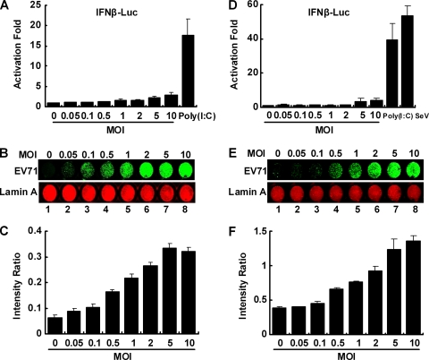
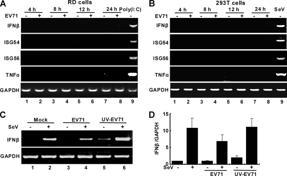
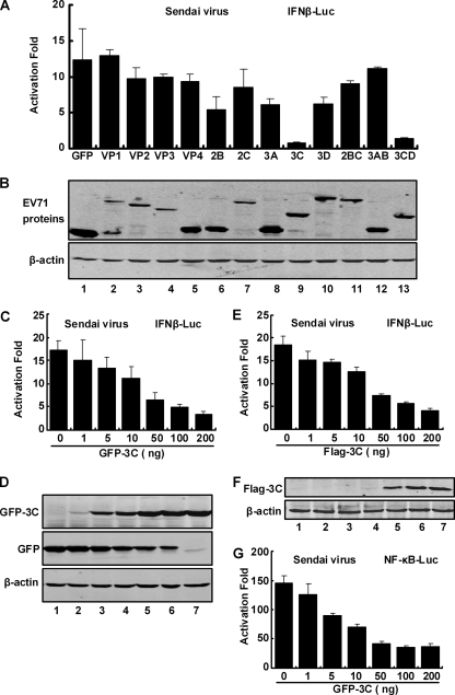
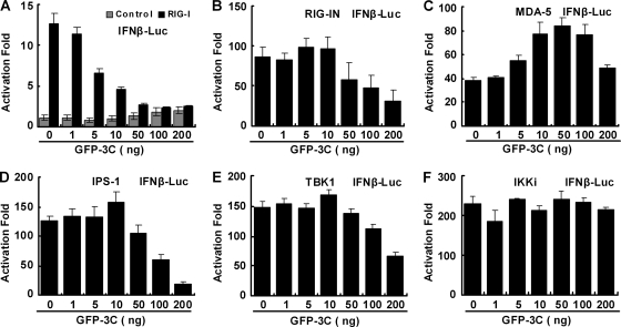
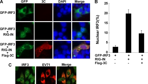
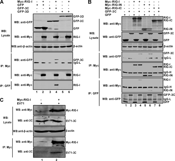
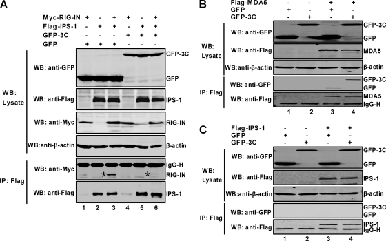
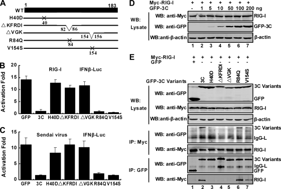
Enterovirus 71 (EV71) is a human pathogen that induces hand, foot, and mouth disease and fatal neurological diseases. Immature or impaired immunity is thought to associate with increased morbidity and mortality. In a murine model, EV71 does not facilitate the production of type I interferon (IFN) that plays a critical role in the first-line defense against viral infection. Administration of a neutralizing antibody to IFN-alpha/beta exacerbates the virus-induced disease. However, the molecular events governing this process remain elusive. Here, we report that EV71 suppresses the induction of antiviral immunity by targeting the cytosolic receptor retinoid acid-inducible gene I (RIG-I). In infected cells, EV71 inhibits the expression of IFN-beta, IFN-stimulated gene 54 (ISG54), ISG56, and tumor necrosis factor alpha. Among structural and nonstructural proteins encoded by EV71, the 3C protein is capable of inhibiting IFN-beta activation by virus and RIG-I. Nevertheless, EV71 3C exhibits no inhibitory activity on MDA5. Remarkably, when expressed in mammalian cells, EV71 3C associates with RIG-I via the caspase recruitment domain. This precludes the recruitment of an adaptor IPS-1 by RIG-I and subsequent nuclear translocation of interferon regulatory factor 3. An R84Q or V154S substitution in the RNA binding motifs has no effect. An H40D substitution is detrimental, but the protease activity associated with 3C is dispensable. Together, these results suggest that inhibition of RIG-I-mediated type I IFN responses by the 3C protein may contribute to the pathogenesis of EV71 infection.


Figures

FIG. 1.
FIG. 1.
(A) Effects of EV71 infection on IFN promoter activation in RD cells. Cells were transfected with pGL3-IFN-β-Luc (100 ng/well and pRL-SV40 (5 ng/well). Cells were then mock infected or infected with EV71 at the indicated multiplicity of infection. As a positive control, cells were transfected with poly(I:C). At 24 h after transfection or infection, cells were harvested to determine luciferase activities. Results are expressed as fold of activation with standard deviations among triplicate samples. (B) EV71 replication on RD cells. Cells were mock infected or infected with EV71 at the indicated multiplicity of infection. At 24 h after infection, cells were stained with antibodies against lamin A and EV71 and subjected to in-cell Western blot analysis as described in Materials and Methods. A representative image is shown, with the green representing EV71 proteins and red representing lamin A, which was used as an internal control. (C) Quantitative analysis of EV71 replication in RD cells. Image signals in panel B were quantified by an Odyssey Infrared Imager (Li-Cor, Lincoln, NE) and expressed as the ratio of EV71 to lamin A. Data are from three independent experiments with standard deviations. (D) Effects of EV71 infection on IFN-β promoter activation in 293T cells. Assays were performed as described for panel A. Poly(I:C) and Sendai virus were used as positive controls. (E) EV71 replication on 293T cells. Assays were done as described for panel B. (F) Quantitative analysis of EV71 replication in 293T cells. Image signals in panel E were quantified and expressed as the ratio of EV71 to lamin A. Data are from three independent experiments with standard deviations.
FIG. 2.
FIG. 2.
EV71 infection does not stimulate the expression of IFN-β, ISG54, ISG56, and TNF-α in RD cells (A) and 293T cells (B). Cells were mock infected or infected with EV71 (MOI of 2). At 4, 8, 12, and 24 h after infection, total RNA extracted from cells was subjected to RT-PCR amplification and electrophoresis for IFN-β, ISG54, ISG56, TNF-α, and GAPDH mRNAs. As controls, mock-infected cells were transfected with poly(I:C) or infected with Sendai virus (SeV) and subjected to RT-PCR analysis. (C) RD cells were mock infected or infected as indicated. At 24 h after infection, total RNA extracted from cells was subjected to RT-PCR analysis for IFN-β and GAPDH mRNAs. (D) RD cells were mock infected or infected as indicated. At 24 h after infection, total RNA extracted from cells was analyzed by quantitative real-time PCR using SYBR Green.
FIG. 3.
FIG. 3.
Inhibition of virus-induced activation of the IFN promoter by EV71 proteins. (A) 293T cells were transfected with pGL3-IFN-β-Luc, a plasmid expressing GFP, and plasmids expressing individual EV71 proteins fused to GFP. A vector plasmid expressing GFP and pRL-SV40 were used as controls. At 24 h after transfection, cells were stimulated with Sendai virus for 24 h, and luciferase activities were measured. Data are representative of three independent experiments with triplicate samples. (B) Expression of EV71-encoded proteins. Lysates of cells in panel A were subjected to Western analysis with antibodies against GFP and β-actin (Sigma, St. Louis, MO). (C) Inhibition of virus-induced IFN-β promoter activation by GFP-3C. 293T cells were transfected with pGL3-IFN-β-Luc, pRL-SV40, and increasing amounts of 3C expression plasmids. Cells were infected with Sendai virus for 24 h and assayed for luciferase activities. Data are expressed as fold of activation with standard deviations among triplicate samples. (D) Expression of GFP-3C. Lysates of cells from panel C were subjected to Western blot analysis with antibodies against GFP and β-actin. (E) Inhibition of virus-induced IFN-β activation by Flag-3C. Assays were done as described for panel C. (F) Expression of Flag-3C. Assays were done as described for panel D with anti-Flag and β-actin antibodies. (G) Inhibition of NF-κB promoter activation by 3C. 293T cells were transfected with NF-κB-Luc, pRL-SV40, and GFP-3C. Cells were infected with Sendai virus for 24 h and assayed for luciferase activities. Data are expressed as fold of activation with standard deviations among triplicate samples.
FIG. 4.
FIG. 4.
Effects of 3C on RIG-I, MDA-5, IPS-1, TBK1, and IKKi. 293T cells were transfected with pGL3-IFN-β-Luc along with plasmid encoding RIG-I (A), RIG-IN (B), MDA5 (C), IPS-1(D), TBK1 (E), or IKKi (F). In addition, pRL-SV40 was included as a control. At 24 or 36 h after transfection, the luciferase activities were measured as described in Materials and Methods. Results are expressed as fold of activation with standard deviations among triplicate samples.
FIG. 5.
FIG. 5.
The 3C protein blocks nuclear translocation of IRF3. (A) 293T cells were cotransfected with GFP-IRF3 alone or along with Myc-RIG-IN and Flag-3C. At 24 h after transfection, cells were processed as described Materials and Methods and visualized under a fluorescence microscope. (B) Quantitation of IRF3 nuclear translocation. A total of 600 GFP-IRF3-positive cells from different fields in panel A were counted. Results are expressed as means ± standard deviations from two independent experiments. (C) RD cells were infected with EV71. At 6 h after infection, cells were stained with anti-EV71 and anti-IRF3 antibodies and visualized by microscope.
FIG. 6.
FIG. 6.
Association of 3C and RIG-I. (A) 3C associates with RIG-I. 293T cells were transfected with plasmids encoding full-length Myc-RIG-I (lanes 2, 4, and 6), GFP (lanes 1 and 2), GFP-3C (lanes 3 and 4), and GFP-3D (lanes 5 and 6). The total amount of DNA was kept constant using the empty vector pcDNA3.1 (lanes 1, 3, and 5). At 24 h after transfection, cell lysates were immunoprecipitated (IP) with antibodies against Myc and GFP (Sigma, St. Louis, MO) separately. Immunoprecipitates and aliquots of cell lysates were then subjected to Western blot (WB) analysis. (B) 3C interacts with the N-terminal domain of RIG-I. 293T cells were transfected with plasmids encoding full-length Myc-RIG-I (lanes 3 and 4), Myc-RIG-IN containing the N-terminal domain (lanes 5 and 6), Myc-RIG-IC containing the C-terminal domain (lanes 7 and 8), GFP-3C (lanes 2, 4, 6, and 8), and GFP (lanes 1, 3, 5, and 7) separately. Immunoprecipitation and Western blot analysis were performed as described for panel A. (C) RD cells, transfected with a plasmid encoding full-length Myc-RIG-I, were infected with EV71 for 20 h, and cell lysates were immunoprecipitated with antibody against Myc. Immunoprecipitates and aliquots of cell lysates were then subjected to Western blot analysis using anti-3C and anti-Myc antibodies.
FIG. 7.
FIG. 7.
3C disrupts the RIG-IN and IPS-1 complex. (A) 293T cells were transfected with plasmid expressing GFP (lanes 1, 2, and 3), Myc-RIG-IN (lanes 1, 3, 4, and 6), GFP-3C (lanes 4, 5, and 6), and Flag-IPS-1 (lanes 2, 3, 5, and 6). Cell lysates were immunoprecipitated (IP) with anti-Flag antibody (Sigma, St. Louis, MO). Immunoprecipitates and aliquots of cell lysates were then subjected to Western blot (WB) analysis with antibodies against Myc, GFP, Flag, and β-actin (Sigma, St. Louis, MO). (B) 293T cells were transfected with GFP (lanes 1 and 3), GFP-3C (lanes 2 and 4), and Flag-MDA5 (lanes 3 and 4). Cell lysates were subjected to immunoprecipitation and Western blot analysis with the indicated antibodies. (C) 293T cells were transfected with plasmids expressing GFP (lanes 1 and 3), GFP-3C (lanes 2 and 4), and Flag-IPS-1 (lanes 3 and 4). Samples were subjected to immunoprecipitation and Western blot analysis with the indicated antibodies.
FIG. 8.
FIG. 8.
Mutational analysis of 3C. (A) Schematic diagrams of 3C variants. The filled bar represents the wild-type 3C of EV71. Other lines represent the 3C mutants as indicated on the figure. H40D bears a substitution of histidine to aspartic acid at amino acid 40. ΔKFRDI has a deletion of amino acids 82 to 86, whereas ΔVGK has a deletion of amino acids 154 to 156. The R84Q variant has a substitution of arginine to glutamine, and V154S has a substitution of valine to serine. (B) Effects of 3C variants on RIG-I-induced IFN β promoter activation. 293T cells were transfected with plasmids encoding RIG-I and IFN-β-Luc along with GFP or GFP-3C variants. pRL-SV40 was included as an internal control. At 36 h transfection, cells were harvested to determine luciferase activities. (C) Effects of 3C variants on Sendai virus-induced IFN-β promoter activation. 293T cells were transfected as described for panel B. At 24 h after transfection, cells were then stimulated with Sendai virus for 24 h, and luciferase activities were measured. Data are expressed as fold of activation with standard deviations among triplicate samples. (D) Effect of 3C on RIG-I expression. 293T cells were transfected with RIG-I along with empty vector or increasing amounts of GFP-3C. At 36 h after transfection, lysates of cells were subjected immunoblot analysis with antibodies against Myc, GFP, and β-actin (Sigma, St. Louis, MO). (E) Interaction of 3C variants with RIG-I. 293T cells were transfected with Myc-RIG-I (all lanes), the GFP-3C variants (as indicated at the top of the panel), and GFP (lane 1) as described Materials and Methods. Cell lysates were immunoprecipitated with anti-Myc antibody and anti-GFP antibody separately. Immunoprecipitates and aliquots of cell lysates were subjected to Western blot analysis with antibodies against Myc, GFP, and β-actin.

Similar articles

Cited by

References

    1. AbuBakar, S., H. Y. Chee, M. F. Al-Kobaisi, J. Xiaoshan, K. B. Chua, and S. K. Lam. 1999. Identification of enterovirus 71 isolates from an outbreak of hand, foot and mouth disease (HFMD) with fatal cases of encephalomyelitis in Malaysia. Virus Res. 61:1-9. - PubMed
    1. AbuBakar, S., I. C. Sam, J. Yusof, M. K. Lim, S. Misbah, N. MatRahim, and P. S. Hooi. 2009. Enterovirus 71 outbreak, Brunei. Emerg. Infect. Dis. 15:79-82. - PMC - PubMed
    1. Alexander, J. P., Jr., L. Baden, M. A. Pallansch, and L. J. Anderson. 1994. Enterovirus 71 infections and neurologic disease—United States, 1977-1991. J. Infect. Dis. 169:905-908. - PubMed
    1. Barral, P. M., J. M. Morrison, J. Drahos, P. Gupta, D. Sarkar, P. B. Fisher, and V. R. Racaniello. 2007. MDA-5 is cleaved in poliovirus-infected cells. J. Virol. 81:3677-3684. - PMC - PubMed
    1. Barral, P. M., D. Sarkar, P. B. Fisher, and V. R. Racaniello. 2009. RIG-I is cleaved during picornavirus infection. Virology 391:171-176. - PMC - PubMed

Publication types

MeSH terms