. 2010 Aug;84(16):8021-32.
doi: 10.1128/JVI.02603-09. Epub 2010 Jun 2.

Chikungunya virus arthritis in adult wild-type mice

Affiliations

Chikungunya virus arthritis in adult wild-type mice

Joy Gardner et al. J Virol. 2010 Aug.

Abstract

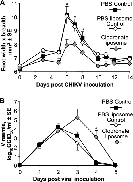
Chikungunya virus is a mosquito-borne arthrogenic alphavirus that has recently reemerged to produce the largest epidemic ever documented for this virus. Here we describe a new adult wild-type mouse model of chikungunya virus arthritis, which recapitulates the self-limiting arthritis, tenosynovitis, and myositis seen in humans. Rheumatic disease was associated with a prolific infiltrate of monocytes, macrophages, and NK cells and the production of monocyte chemoattractant protein 1 (MCP-1), tumor necrosis factor alpha (TNF-alpha), and gamma interferon (IFN-gamma). Infection with a virus isolate from the recent Reunion Island epidemic induced significantly more mononuclear infiltrates, proinflammatory mediators, and foot swelling than did an Asian isolate from the 1960s. Primary mouse macrophages were shown to be productively infected with chikungunya virus; however, the depletion of macrophages ameliorated rheumatic disease and prolonged the viremia. Only 1 microg of an unadjuvanted, inactivated, whole-virus vaccine derived from the Asian isolate completely protected against viremia and arthritis induced by the Reunion Island isolate, illustrating that protection is not strain specific and that low levels of immunity are sufficient to mediate protection. IFN-alpha treatment was able to prevent arthritis only if given before infection, suggesting that IFN-alpha is not a viable therapy. Prior infection with Ross River virus, a related arthrogenic alphavirus, and anti-Ross River virus antibodies protected mice against chikungunya virus disease, suggesting that individuals previously exposed to Ross River virus should be protected from chikungunya virus disease. This new mouse model of chikungunya virus disease thus provides insights into pathogenesis and a simple and convenient system to test potential new interventions.


Figures

FIG. 1.
FIG. 1.
Disease and virus replication. (A) Pictures of feet at the time (day 7) of peak swelling after inoculation of medium (control) or the Asian or the Reunion isolate of CHIKV. (B) Perimetatarsal foot swelling over time after inoculations described above (A) (n = 6 to 12 feet per group). Peak swelling (day 7 for Reunion isolates and day 8 for the Asian isolate) is indicated (*) and was ∼75% higher for the Reunion Island isolate (P = 0.024). (C) Peripheral blood viremia (n = 6). (D) Virus titers in the foot (n = 6 to 10 mice per group). (E) Virus titers in quadriceps muscle, spleen, inguinal lymph nodes, and liver (n = 3 to 6 mice per group). (F) Replication (*, P = 0.03) and induction of CPE (*, P = 0.021 and 0.029) following infection of MEFs by the two CHIKV isolates (MOI of 0.1) (means of data from four replicates).
FIG. 2.
FIG. 2.
Histology and immunocytochemistry of feet from control mice or mice infected with the Asian or Reunion Island CHIKV isolate day 7 postinfection. The row labeled “Foot” shows subcutaneous edema (*), and foci of inflammatory cell infiltrates in muscle tissue (arrows) are indicated (bars, 1 mm). B, bone; M, muscle. The row labeled “Joint” shows that synovial membranes (arrowheads) were disrupted and contained mononuclear cell infiltrates (bars, 100 μm). SS, synovial space. The row labeled “Synovium” shows that the normal cohesive cells lining the synovial membrane (control) (arrows) were absent in infected mice, sometimes replaced by moderate amounts of fibrinous exudate (columns labeled “Asian” and “Reunion”) (arrows). The basement membrane was always visible (arrowheads). Numerous inflammatory cells, composed mainly of large mononuclear cells, have infiltrated the underlying connective tissue in infected mice (bars, 10 μm). The row labeled “Tendon” shows marked tenosynovitis in CHIKV-infected mice, with inflammatory cells present in the tendon capsule and sometimes in the distended space (arrowhead) between the capsule and the tendon (bars, 100 μm). T, tendon. In the row labeled “Muscle,” extensive inflammatory infiltrate can be seen in CHIKV-infected mice, which was more pronounced in mice infected with the Reunion Island isolate (bars, 100 μm). The row labeled “F4/80” shows metatarsal areas stained with the F4/80 macrophage-specific antibody (bars, 1 mm).
FIG. 3.
FIG. 3.
Clodronate depletion of macrophages. Mice (n = 3 to 4 per group) were treated with clodronate liposomes, control liposomes, or PBS and were infected on the next day with the Reunion Island isolate of CHIKV. (A) Foot swelling showing significant differences on the indicated (*) days (day 6, P = 0.002; day 7, P = 0.01; day 8, P = 0.045). (B) Mean viremia in the same mice described above (A). Viremia was significantly different between the clodronate liposome and control liposome groups on day 4 (*) (P = 0.037).
FIG. 4.
FIG. 4.
Infection of macrophages. (A) Splenocytes were infected with CHIKV carrying GFP (MOI of 2) for 24 h, stained with anti-F4/80-PE, and analyzed by FACS. The area of GFP+ F4/80+ cells is indicated by a box with dotted lines. (B) Cultured adherent cells from spleen (2 × 10⁴ cells/well) (>85% F4/80+) were infected with the indicated MOIs of the Reunion Island isolate. Cells were then washed extensively, and supernatants were then assayed for viral titers at 0, 1, 2, and 3 days. The means of data from three replicates are shown.
FIG. 5.
FIG. 5.
Inflammatory mediator profiles. (A) Mice were infected (n = 4 to 8 per group) with Reunion Island or Asian CHIKV isolates, and on the indicated days, serum was analyzed for the indicated cytokines/chemokine. An asterisk indicates where levels were significantly different (P = 0.007 for TNF-α; P = 0.001, 0.006, 0.004, and 0.004 for days 1, 2, 4, and 7, respectively for MCP-1; P = 0.007 for IFN-γ; and P = 0.015 for IFN-α/β). (B) Feet (n = 4) taken 6 days postinfection were analyzed for cytokine/chemokine mRNA levels using real-time RT-PCR. mRNA levels for Reunion Island and Asian isolate-infected mice were significantly different for MCP-1 (P = 0.019) and IFN-γ (P = 0.017).
FIG. 6.
FIG. 6.
Vaccination and CHIKV challenge. (A) Fifty percent endpoint IgG1 and IgG2c titers 5 weeks after infection (n = 6) with the indicated virus isolate or vaccination with different doses of purified inactivated CHIKV vaccine (n = 3 per group). (B) Five weeks after vaccination, mice were challenged with the Reunion Island isolate, and the level of viremia was determined over 6 days. Mock vaccination was done with medium. (C) Foot swelling of the animals shown in B (n = 6 feet per group).
FIG. 7.
FIG. 7.
Cross-protection with RRV. (A) Mice were infected with RRV or mock infected (n = 6 per group) and 4.5 months later were infected with the Reunion Isolate of CHIKV, and CHIKV viremia was measured (an asterisk indicates significant differences [P = 0.029 and 0.005]). (B) CHIKV-induced foot swelling of mice that had received serum from naïve mice or mice previously infected with RRV or the Reunion Island isolate of CHIKV. (C) ELISA using serum from the RRV-infected mice described above (A) prior to CHIKV infection, with titers determined on purified inactivated CHIKV. (D) IFN-γ ELISPOT assay using splenocytes from a separate group of mice infected 4.5 weeks previously with the indicated virus and using purified inactivated CHIKV as an antigen.

References

    1. Aaskov, J., L. Williams, and S. Yu. 1997. A candidate Ross River virus vaccine: preclinical evaluation. Vaccine 15:1396-1404. - PubMed
    1. Akahata, W., Z. Y. Yang, H. Andersen, S. Sun, H. A. Holdaway, W. P. Kong, M. G. Lewis, S. Higgs, M. G. Rossmann, S. Rao, and G. J. Nabel. 2010. A virus-like particle vaccine for epidemic Chikungunya virus protects nonhuman primates against infection. Nat. Med. 16:334-338. - PMC - PubMed
    1. Alsharifi, M., M. Lobigs, M. M. Simon, A. Kersten, K. Muller, A. Koskinen, E. Lee, and A. Mullbacher. 2006. NK cell-mediated immunopathology during an acute viral infection of the CNS. Eur. J. Immunol. 36:887-896. - PubMed
    1. Anraku, I., V. V. Mokhonov, P. Rattanasena, E. I. Mokhonova, J. Leung, G. Pijlman, A. Cara, W. A. Schroder, A. A. Khromykh, and A. Suhrbier. 2008. Kunjin replicon-based simian immunodeficiency virus gag vaccines. Vaccine 26:3268-3276. - PMC - PubMed
    1. Antalis, T. M., M. La Linn, K. Donnan, L. Mateo, J. Gardner, J. L. Dickinson, K. Buttigieg, and A. Suhrbier. 1998. The serine proteinase inhibitor (serpin) plasminogen activation inhibitor type 2 protects against viral cytopathic effects by constitutive interferon alpha/beta priming. J. Exp. Med. 187:1799-1811. - PMC - PubMed

Publication types

MeSH terms