. 2010 Aug;84(16):8262-74.
doi: 10.1128/JVI.00385-10. Epub 2010 Jun 2.

Accessory protein 5a is a major antagonist of the antiviral action of interferon against murine coronavirus

Affiliations

Accessory protein 5a is a major antagonist of the antiviral action of interferon against murine coronavirus

Cheri A Koetzner et al. J Virol. 2010 Aug.

Abstract

The type I interferon (IFN) response plays an essential role in the control of in vivo infection by the coronavirus mouse hepatitis virus (MHV). However, in vitro, most strains of MHV are largely resistant to the action of this cytokine, suggesting that MHV encodes one or more functions that antagonize or evade the IFN system. A particular strain of MHV, MHV-S, exhibited orders-of-magnitude higher sensitivity to IFN than prototype strain MHV-A59. Through construction of interstrain chimeric recombinants, the basis for the enhanced IFN sensitivity of MHV-S was found to map entirely to the region downstream of the spike gene, at the 3' end of the genome. Sequence analysis revealed that the major difference between the two strains in this region is the absence of gene 5a from MHV-S. Creation of a gene 5a knockout mutant of MHV-A59 demonstrated that a major component of IFN resistance maps to gene 5a. Conversely, insertion of gene 5a, or its homologs from related group 2 coronaviruses, at an upstream genomic position in an MHV-A59/S chimera restored IFN resistance. This is the first demonstration of a coronavirus gene product that can protect that same virus from the antiviral state induced by IFN. Neither protein kinase R, which phosphorylates eukaryotic initiation factor 2, nor oligoadenylate synthetase, which activates RNase L, was differentially activated in IFN-treated cells infected with MHV-A59 or MHV-S. Thus, the major IFN-induced antiviral activities that are specifically inhibited by MHV, and possibly by other coronaviruses, remain to be identified.


Figures

FIG. 1.
FIG. 1.
Differing IFN sensitivities of MHV-A59 and MHV-S. Mouse L2 cell monolayers were treated with the indicated doses of IFN-α for 24 h and then infected at a multiplicity of 2.0 PFU per cell. Virus was harvested at 18 h postinfection, and infectious titers were determined by plaque assay on L2 cells. Open and shaded symbols represent results from two separate experiments. In the absence of IFN, the infectious titers were 6.3 × 10⁷ and 6.7 × 10⁷ PFU/ml for MHV-A59 and 1.5 × 10⁷ and 1.0 × 10⁷ PFU/ml for MHV-S. At a dose of 1,000 U/ml IFN, the infectious titers were 7.2 × 10⁶ and 7.3 × 10⁶ PFU/ml for MHV-A59 and 1.9 × 10⁴ and 6.4 × 10³ PFU/ml for MHV-S.
FIG. 2.
FIG. 2.
Comparison of the genomic compositions of MHV-A59 and MHV-S. For each strain, the region downstream of replicase gene 1 is shown. Rectangles represent ORFs for structural and accessory proteins; dotted lines indicate pseudogenes. In each MHV-S ORF, vertical lines indicate amino acid residues that are nonidentical to those of MHV-A59; note that this representation accentuates differences between the ORFs of the two strains (see Table 1). Open circles represent TRSs; the filled circle marks the nonfunctional mutant TRS upstream of MHV-S gene 4. The solid triangle highlights the deletion of gene 5a in MHV-S.
FIG. 3.
FIG. 3.
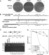
Mapping of the IFN resistance of MHV-A59. (A) Differential effect of IFN on the sizes of plaques formed by MHV-A59 and MHV-S. L2 cells were treated with 0, 10, or 100 U/ml IFN-α for 24 h prior to the performance of standard plaque assays. Monolayers were stained with neutral red at 48 h postinfection and photographed 4 h later. (B) Targeted recombination between MHV-S genomic RNA and synthetic donor RNA transcribed from pMH54, which contains all of the MHV-A59 genes downstream of the HE gene (24). Progeny obtained from infected and transfected cells were screened on L2 cells that had been pretreated with 10 or 100 U/ml IFN-α, and candidate recombinants were isolated as viruses that formed large plaques. (C) Analysis of eight candidate recombinants. In the schematic, vertical lines in each MHV-S ORF indicate amino acid residues that are nonidentical to those of MHV-A59. Note that primers (arrows) are not drawn to scale; the two primer hybridization sites are identical between MHV-A59 and MHV-S. RT-PCR products crossing the gene 5a region were analyzed by agarose gel electrophoresis; the sizes (base pairs) of markers in the first lane are indicated to the left of the gel. Denoted beneath the gel are the five independent groups (a to e) into which the recombinants were found to fall, based on sequencing of the crossover sites in the HE gene. (D) IFN dose responses of independent recombinants 4 and 8, compared to MHV-A59 and MHV-S. Mouse L2 cell monolayers were treated with IFN-α for 24 h and then infected at a multiplicity of 2.0 PFU per cell. Virus was harvested at 18 h postinfection, and infectious titers were determined by plaque assay on L2 cells.
FIG. 4.
FIG. 4.
Mapping of the IFN sensitivity of MHV-S. (A) Schematic comparison of the genomes of MHV-A59, MHV-S, and the chimera MHV-A59/S; the latter was constructed by targeted RNA recombination as described in Materials and Methods. MHV-A59/S contains, in an otherwise MHV-A59 background, all of the MHV-S sequence downstream of the S gene (including nonfunctional TRS4 of MHV-S). The 3′ untranslated region of the chimeric virus corresponds to that of MHV-A59 but differs from the MHV-S 3′ untranslated region by only 1 nt. (B) IFN dose response of MHV-A59/S, compared to those of MHV-A59 and MHV-S. Mouse L2 cell monolayers were treated with IFN-α for 24 h and then infected at a multiplicity of 2.0 PFU per cell. Virus was harvested at 18 h postinfection, and infectious titers were determined by plaque assay on L2 cells. Open and shaded symbols represent results from two separate experiments.
FIG. 5.
FIG. 5.
IFN sensitivity of the MHV-A59 gene 5a knockout mutant. (A) Construction of the 5a-KO mutant. Shown above the schematic of the MHV-A59 genome is the wild-type sequence of the gene 4-gene 5a junction. TRS5 is located within ORF 4, 32 nt upstream of the sequence segment shown. Indicated below the genome are four point mutations made to eliminate the start codon of ORF 5a and to replace the fourth codon with a stop codon. Mutations were constructed in donor RNA transcription vector pMH54 (24) and incorporated into MHV-A59 via targeted RNA recombination as described in Materials and Methods. (B) IFN dose response of the 5a-KO mutant, compared to those of MHV-A59 and MHV-S. Mouse L2 cell monolayers were treated with IFN-α for 24 h and then infected at a multiplicity of 2.0 PFU per cell. Virus was harvested at 18 h postinfection, and infectious titers were determined by plaque assay on L2 cells. Open and shaded symbols represent results from two separate experiments. (C) Western blot assays of lysates from 17Cl1 cells that were either mock infected or infected with wild-type MHV or the 5a-KO mutant at a multiplicity of 1.0 or 0.2 PFU per cell. Blots were probed with polyclonal anti-MHV E antiserum (25) or with monoclonal anti-N antibody J.3.3.
FIG. 6.
FIG. 6.
IFN sensitivities of chimeras of MHV-A59 containing the MHV-S N gene in the presence or absence of the gene 5a knockout. (A) Schematics of the 3′ ends of mutant genomes, compared to those of MHV-A59 and MHV-S. In each MHV-S ORF, vertical lines indicate amino acid residues that are nonidentical to those of MHV-A59; X denotes the 5a-KO mutations shown in detail in Fig. 5A. The N chimera mutant (and the 5a-KO+N chimera mutant) contain all of the amino acid residues of the MHV-S N protein that differ from those of the MHV-A59 N protein, except for V321A; additionally, the carboxy-terminal residue of the M protein (T228) is changed to its MHV-S counterpart (I228). Mutants were constructed by targeted RNA recombination as described in Materials and Methods. (B) IFN dose responses of chimeras, compared to those of MHV-A59, MHV-S, and the 5a-KO mutant. Mouse L2 cell monolayers were treated with IFN-α for 24 h and then infected at a multiplicity of 2.0 PFU per cell. Virus was harvested at 18 h postinfection, and infectious titers were determined by plaque assay on L2 cells. Open and shaded symbols represent results from two separate experiments.
FIG. 7.
FIG. 7.
Effect of substitution of gene 5a in the gene 2 region of the MHV-A59/S chimera. (A) Schematic comparison of the genomes of MHV-A59, the MHV-A59Δ2a-HE mutant, the MHV-A59/S chimera, and mutants in which the 2a-HE region of the MHV-A59/S chimera was replaced with gene 5a of MHV-A59 or the homolog of gene 5a from BCoV or HCoV-HKU1. Deletion and substitution mutants were constructed by targeted RNA recombination as described in Materials and Methods. (B) Amino acid sequence alignment of the MHV-A59 5a protein and its homologs from BCoV (12.7-kDa protein) and HCoV-HKU1 (ORF 4 product). The GenBank accession numbers of the sequences shown are as follows: MHV-A59, AY700211; BCoV, U00735; HCoV-HKU1, AY597011. Circles denote residues that are conserved among all group 2a coronaviruses. (C and D) Relative IFN resistances of gene 5a substitution mutants. The graph (C) shows the IFN dose response of the MHV-A59/S+5a(MHV) substitution mutant, compared to MHV-A59 and the MHV-A59/S chimera. Mouse L2 cell monolayers were treated with IFN-α for 24 h and then infected at a multiplicity of 2.0 PFU per cell. Virus was harvested at 18 h postinfection, and infectious titers were determined by plaque assay on L2 cells. Open and shaded symbols represent results from two separate experiments. Histograms (D) show the relative infectious titers (compared to untreated samples) for all three of the 5a homolog substitution mutants, compared to those of the control viruses, at an IFN-α dose of 100 U/ml. Each value is the mean of three replicate samples ± the standard deviation.
FIG. 8.
FIG. 8.
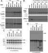
Status of the PKR/eIF2α and OAS/RNase L antiviral systems in MHV-infected cells. Mouse L2 cell monolayers were left untreated or treated with 100 U/ml IFN-α for 24 h. Monolayers were then mock infected or infected with MHV-A59, MHV-S, or the MHV-A59/S chimera at a multiplicity of 2.0 PFU per cell. Cell lysates were prepared at 6 or 10 h postinfection. One untreated, mock-infected sample was treated with 0.5 mM sodium m-arsenite for 30 min prior to harvesting to provide a positive control for eIF2α phosphorylation (10). Lysates were analyzed by Western blot assays probed with antibodies specific for PKR (A) or for total or phosphorylated eIF2α (B). (C) For one set of samples, cells were harvested at 10 h postinfection and total cellular RNA was purified and analyzed by electrophoresis in a 1% agarose gel stained with ethidium bromide. The sizes (base pairs) of double-stranded DNA markers in the first lane are indicated to the left of the gel; the positions of the 28S and 18S rRNAs are shown at the right. (D) In a separate experiment, total cellular RNA was purified at 10 h postinfection from IFN-treated cells that had been mock infected or infected with MHV-A59, MHV-S, the MHV-A59/S chimera, encephalomyocarditis virus (EMCV), reovirus type 3, or mengovirus. RNA was separated by electrophoresis in a 1.2% agarose gel containing formaldehyde and analyzed by Northern blotting using a probe specific for the 18S rRNA.

References

    1. Abraham, S., T. E. Kienzle, W. E. Lapps, and D. A. Brian. 1990. Sequence and expression analysis of potential nonstructural proteins of 4.9, 4.8, 12.7, and 9.5 kDa encoded between the spike and membrane protein genes of the bovine coronavirus. Virology 177:488-495. - PMC - PubMed
    1. Budzilowicz, C. J., and S. R. Weiss. 1987. In vitro synthesis of two polypeptides from a nonstructural gene of coronavirus mouse hepatitis virus strain A59. Virology 157:509-515. - PMC - PubMed
    1. Cervantes-Barragán, L., U. Kalinke, R. Züst, M. König, B. Reizis, C. López-Macías, V. Thiel, and B. Ludewig. 2009. Type I IFN-mediated protection of macrophages and dendritic cells secures control of murine coronavirus infection. J. Immunol. 182:1099-1106. - PubMed
    1. Cervantes-Barragan, L., R. Zust, F. Weber, M. Spiegel, K. S. Lang, S. Akira, V. Thiel, and B. Ludewig. 2007. Control of coronavirus infection through plasmacytoid dendritic-cell-derived type I interferon. Blood 109:1131-1137. - PMC - PubMed
    1. Cheley, S., V. L. Morris, M. J. Cupples, and R. Anderson. 1981. RNA and peptide homology among murine coronaviruses. Virology 115:310-321. - PMC - PubMed

Publication types

MeSH terms