. 2010 Oct;91(Pt 10):2542-52.
doi: 10.1099/vir.0.023085-0. Epub 2010 Jun 2.

Vaccination with murid herpesvirus-4 glycoprotein B reduces viral lytic replication but does not induce detectable virion neutralization

Affiliations

Vaccination with murid herpesvirus-4 glycoprotein B reduces viral lytic replication but does not induce detectable virion neutralization

Janet S May et al. J Gen Virol. 2010 Oct.

Abstract

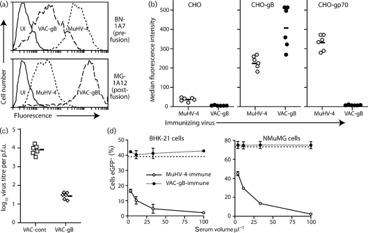
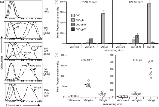
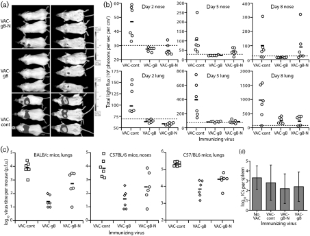
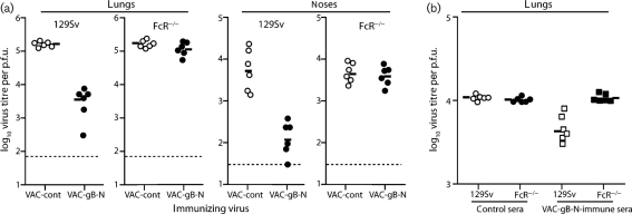
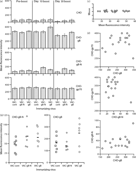
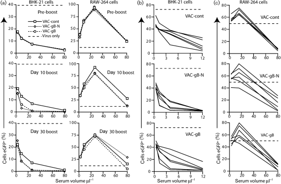
Herpesviruses characteristically disseminate from immune hosts. Therefore in the context of natural infection, antibody neutralizes them poorly. Murid herpesvirus-4 (MuHV-4) provides a tractable model with which to understand gammaherpesvirus neutralization. MuHV-4 virions blocked for cell binding by immune sera remain infectious for IgG-Fc receptor(+) myeloid cells, so broadly neutralizing antibodies must target the virion fusion complex - glycoprotein B (gB) or gH/gL. While gB-specific neutralizing antibodies are rare, its domains I+II (gB-N) contain at least one potent neutralization epitope. Here, we tested whether immunization with recombinant gB presenting this epitope could induce neutralizing antibodies in naive mice and protect them against MuHV-4 challenge. Immunizing with the full-length gB extracellular domain induced a strong gB-specific antibody response and reduced MuHV-4 lytic replication but did not induce detectable neutralization. gB-N alone, which more selectively displayed pre-fusion epitopes including neutralization epitopes, also failed to induce neutralizing responses, and while viral lytic replication was again reduced this depended completely on IgG Fc receptors. gB and gB-N also boosted neutralizing responses in only a minority of carrier mice. Therefore, it appears that neutralizing epitopes on gB are intrinsically difficult for the immune response to target.


Figures

Fig. 1.
Fig. 1.
Expression of gB epitopes by vaccinia virus-expressed gB and gB-N. (a) BHK-21 cells were left uninfected (UI) or infected (1 p.f.u. per cell, 14 h) with either MuHV-4 or a vaccinia virus expressing a GPI-linked gB extracellular domain (VAC-gB). Cell surface gB expression was analysed by flow cytometric staining of pre-fusion gB with mAb BN-1A7, and post-fusion gB with MG-1A12. (b) C57BL/6 mice were infected i.p. with either MuHV-4 or VAC-gB. Sera collected 2 weeks later were used to stain CHO cells either untransfected or transfected with gp70 or GPI-linked gB. Each point shows the median fluorescence intensity for a 1/500 serum dilution from one mouse. The bars show mean values. CHO-gB staining was significantly greater after VAC-gB infection than after MuHV-4 infection (P<0.01 by Student's two-tailed t-test). In this and subsequent experiments, consistent results were obtained with least two further serum dilutions. (c) Mice were immunized i.p. with either VAC-gB or a control, and 1 month later infected i.n. with MuHV-4. Infectious MuHV-4 in lungs was plaque assayed at 5 days post-challenge. Each point shows the titre of one mouse. The bars show geometric means. (d) Sera from mice infected with either VAC-gB or MuHV-4 were used to neutralize eGFP+ MuHV-4 for BHK-21 cell and NMuMG cell infections. Infection was assayed by flow cytometry for eGFP expression 18 h after exposure to virus/antibody. Each point shows mean±sd of triplicate infections. The dashed lines show infection with virus alone.
Fig. 2.
Fig. 2.
Primary antibody responses to gB vaccines in C57BL/6 and BALB/c mice. (a) BHK-21 cells were left uninfected (UI) or infected (1 p.f.u. per cell, 14 h) with vaccinia virus expressing either the full-length gB extracellular domain (VAC-gB), just its N-terminal domains (VAC-gB-N), or a control insert (VAC-cont). gB expression was analysed by flow cytometry. mAbs BN-6E1 and SC-9E8 recognize neutralizing epitopes on pre-fusion gB. MG-1A12 recognizes a post-fusion epitope that is not contained within gB-N. (b) Sera from mice infected i.p. 1 month previously with VAC-gB, VAC-gB-N or a control virus were pooled from six mice per group and assayed for reactivity to gB, gB-N and gp70 (as a negative control) by flow cytometry of transfected CHO cells. Bars show mean±sd values. For both C57BL/6 and BALB/c mice, VAC-gB-N primed CHO-gB-N-specific responses significantly better than did VAC-gB, and VAC-gB primed CHO-gB-N-specific responses significantly better than did VAC-gB-N (P<0.0001 by Student's two-tailed t-test). (c) Individual sera from the C57BL/6 mice in (b) were assayed for reactivity to gB and gB-N. Each point shows staining of the relevant CHO cells by serum from one mouse. The bars show mean values. VAC-gB-primed mice showed significantly stronger staining of both CHO-gB (P<0.0001) and CHO-gB-N (P<0.01), as did VAC-gB-N-primed mice (P<0.0001 by Student's two-tailed t-test). Each group also showed significantly stronger staining of the cognate form of gB (P<0.0001).
Fig. 3.
Fig. 3.
Protection against MuHV-4 lytic replication by vaccination with gB. (a) BALB/c mice were immunized i.p. with vaccinia viruses expressing gB, gB-N or an irrelevant insert (VAC-cont), and 1 month later challenged i.n. (104 p.f.u.) with luciferase+ MuHV-4. Viral replication in lungs and noses was monitored by luciferin injection and CCD camera scanning. A representative image is shown of mice 5 days post-challenge. (b) Mice were immunized then infected with luciferase+ MuHV-4 as in (a). Each point shows the luciferase signal for one mouse. The bars show mean values. Dashed lines show the lower limits of signal detection. Priming with either VAC-gB or VAC-gB-N significantly reduced luciferase signals in lungs at all time points (P<0.03 by Student's two-tailed t-test). Luciferase signals in noses were also significantly reduced in VAC-gB-primed mice at days 2 (P<0.01) and 5 (P<0.04), and in VAC-gB-N-primed mice at day 2 (P<0.01). (c) BALB/c or C57BL/6 mice were immunized i.p. with vaccinia virus recombinants then challenged i.n. with MuHV-4 as in (b), but with luciferase MuHV-4. Lungs and noses were titrated for infectious virus at 6 days post-infection by plaque assay. Each point shows the titre of one mouse. The bars show geometric means. Both BALB/c (P<0.04) and C57BL/6 mice (P<0.01) lung titres were significantly reduced by priming with either VAC-gB or VAC-gB-N. C57BL/6 mice nose titres were also reduced, although this was only statistically significant for VAC-gB (P<0.01). (d) Latent virus loads in spleens were measured by infectious centre assay at 2 months post-challenge of vaccinated C57BL/6 mice. The bars show mean±sd titres of five mice per group.
Fig. 4.
Fig. 4.
gB-primed sera fail to neutralize MuHV-4. (a) BALB/c mice were primed with vaccinia virus expressing gB, gB-N or a control insert. After 1 month, sera pooled from six mice per group were tested for their capacity to reduce BHK-21 or RAW-264 cell infections by eGFP+ MuHV-4, as assayed by flow cytometry 16 h after exposure to virus. Neutralization by sera pooled from MuHV-4-infected mice (Imm ser) is also shown. Dashed lines show the level of infection by virus alone. (b) In a similar protocol to (a), VAC-gB-primed and VAC-gB-N-primed sera, each pooled from six C57BL/6 mice, showed no significant MuHV-4 neutralization. (c) Sera from the mice in (b) were tested individually for neutralization of BHK-21 cell infection. Again none was observed. The dashed line shows neutralization by pooled, MuHV-4-immune sera. (d) Sera pooled from unprimed or VAC-primed mice (n=7) were collected either before or 16 days after MuHV-4 challenge and used to neutralize MuHV-4 virions for BHK-21 cell infection. Each line shows mean±sem results. No significant difference was observed between VAC-gB-N-primed and control mice.
Fig. 5.
Fig. 5.
Protection of naive mice and induction of MuHV-4 neutralization by vaccination with a gHL fusion protein. (a) 129Sv×C57BL/6 FcRγ−/−FcγRII−/− mice (FcR−/−) or 129Sv controls were primed i.p. with vaccinia virus recombinants (105 p.f.u.) and 1 month later challenged i.n. with MuHV-4 (104 p.f.u.). Infectious virus titres in lungs and noses were measured by plaque assay 6 days later. Each point shows the result for one mouse. The bars show geometric means. VAC-gB significantly reduced virus replication in lungs (P<0.00001) and noses (P<0.04) of 129SV but not FcR−/− mice. (b) Sera were pooled from mice (n=10) 3 months after i.p. infection with VAC-gB-N or VAC-cont, then given i.p. to 129Sv or FcR−/− mice (500 μl per mouse) at the same time as i.n. MuHV-4 (5000 p.f.u.). Lungs were titrated for infectious virus by plaque assay 5 days later. Each point shows the result for one mouse, and the bars show geometric means. gB-N-immune sera significantly reduced virus titres in 129Sv mice (P<0.0001 by Student's two-tailed t-test) but not in FcR−/− mice (P=0.86).
Fig. 6.
Fig. 6.
Boosting gB-specific antibody responses by post-exposure vaccination of MuHV-4 carrier mice. (a) C57BL/6 mice were infected i.n. with MuHV-4 and 3 months later boosted i.p. with vaccinia virus expressing either the full-length gB extracellular domain (VAC-gB), just its N-terminal half (VAC-gB-N) or a control insert (VAC-cont). Sera were taken before vaccinia infection (pre-boost), 10 and 30 days later, and tested for gB reactivity by flow cytometric staining of CHO cells expressing gB or gB-N. CHO cells either untransfected or transfected with the MuHV-4 gp70 provided staining controls. Bars show mean±sd fluorescence intensities for sera pooled from six mice each. VAC-gB-N and VAC-gB both significantly increased CHO-gB-N staining at days 10 and 30 compared with VAC-cont (P<0.0001 by Student's two-tailed t-test). The increase with VAC-gB-N was significantly greater than with VAC-gB (P<0.0001). VAC-gB but not VAC-gB-N significantly increased CHO-gB staining at days 10 and 30 compared with VAC-cont (P<0.0001). (b) Sera from the individual day 30 boosted mice in (a) were assayed for CHO-gB and CHO-gB-N reactivity. Although boosting increased staining, this did not reach statistical significance (P>0.05) because of wide individual variation. Each point shows an individual mouse and the bars show geometric means. (c) C57BL/6 MuHV-4 carrier mice (3 months post-infection) were analysed by flow cytometry for serum binding to CHO-gB-N cells. Each point shows the result for one mouse. (d) A separate set of MuHV-4-infected C57BL/6 mice were compared for serum reactivity to CHO-gB, CHO-gB-N and CHO-gp70 cells. Each point shows the result for one mouse. There was no obvious correlation for individual mice between the responses to each target.
Fig. 7.
Fig. 7.
Boosting gB-specific neutralizing antibody responses by post-exposure vaccination of MuHV-4 carrier mice. (a) Sera taken pre-boosting, 10 or 30 days post-boosting with vaccinia viruses expressing the full-length gB extracellular domain (VAC-gB), its N-terminal half (VAC-gB-N) or an irrelevant protein (VAC-cont), were pooled from groups of six C57BL/6 mice each. eGFP+ MuHV-4 virions were incubated with dilutions of the pooled sera and then used to infect either BHK-21 cells (0.3 p.f.u. per cell) or RAW-264 cells (3 p.f.u. per cell). Infected cells were enumerated 16 h later by flow cytometry. Dashed lines show infection with untreated virus. Each point gives the result for 10 000 cells. Thus by χ2 test, the visible reductions in infection with VAC-gB or VAC-gB boosting were all highly significant (P<0.0001). The day 30 RAW-264 cell infection differences were not considered significant as they were not maintained over more than 1 serum dilution. (b) Individual sera taken at day 30 post-boosting were analysed for neutralization of BHK-21 cell infection as in (a). With 4 μl serum, both the VAC-gB (P<0.05) and VAC-gB-N boosted groups showed a significant reduction in infection (P<0.03 by Student's two-tailed t-test) compared with the control group. With 1.3 μl serum, only the VAC-gB-N boosted group showed a reduction (P<0.03). (c) Sera taken at day 30 post-boosting were analysed for neutralization of RAW-264 cell infection (3 p.f.u. per cell). The boosted groups showed a wider distribution than the unboosted, but no significant reduction.

Similar articles

Cited by

References

    1. Adler, H., Messerle, M., Wagner, M. & Koszinowski, U. H. (2000). Cloning and mutagenesis of the murine gammaherpesvirus 68 genome as an infectious bacterial artificial chromosome. J Virol 74, 6964–6974. - PMC - PubMed
    1. Akula, S. M., Pramod, N. P., Wang, F. Z. & Chandran, B. (2001). Human herpesvirus 8 envelope-associated glycoprotein B interacts with heparan sulfate-like moieties. Virology 284, 235–249. - PubMed
    1. Bennett, N. J., May, J. S. & Stevenson, P. G. (2005). Gamma-herpesvirus latency requires T cell evasion during episome maintenance. PLoS Biol 3, e120. - PMC - PubMed
    1. Burton, D. R., Stanfield, R. L. & Wilson, I. A. (2005). Antibody vs. HIV in a clash of evolutionary titans. Proc Natl Acad Sci U S A 102, 14943–14948. - PMC - PubMed
    1. Cranage, M. P., Kouzarides, T., Bankier, A. T., Satchwell, S., Weston, K., Tomlinson, P., Barrell, B., Hart, H., Bell, S. E. & other authors (1986). Identification of the human cytomegalovirus glycoprotein B gene and induction of neutralizing antibodies via its expression in recombinant vaccinia virus. EMBO J 5, 3057–3063. - PMC - PubMed

Publication types

LinkOut - more resources