

In situ molecular diagnosis and histopathological characterization of enteroadherent Enterococcus hirae infection in pre-weaning-age kittens
- PMID: 20519483
- PMCID: PMC2916556
- DOI: 10.1128/JCM.00916-09
In situ molecular diagnosis and histopathological characterization of enteroadherent Enterococcus hirae infection in pre-weaning-age kittens
Abstract
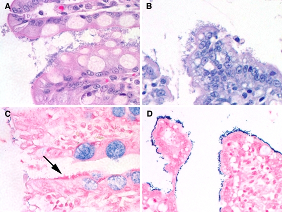
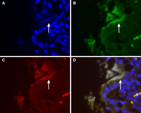
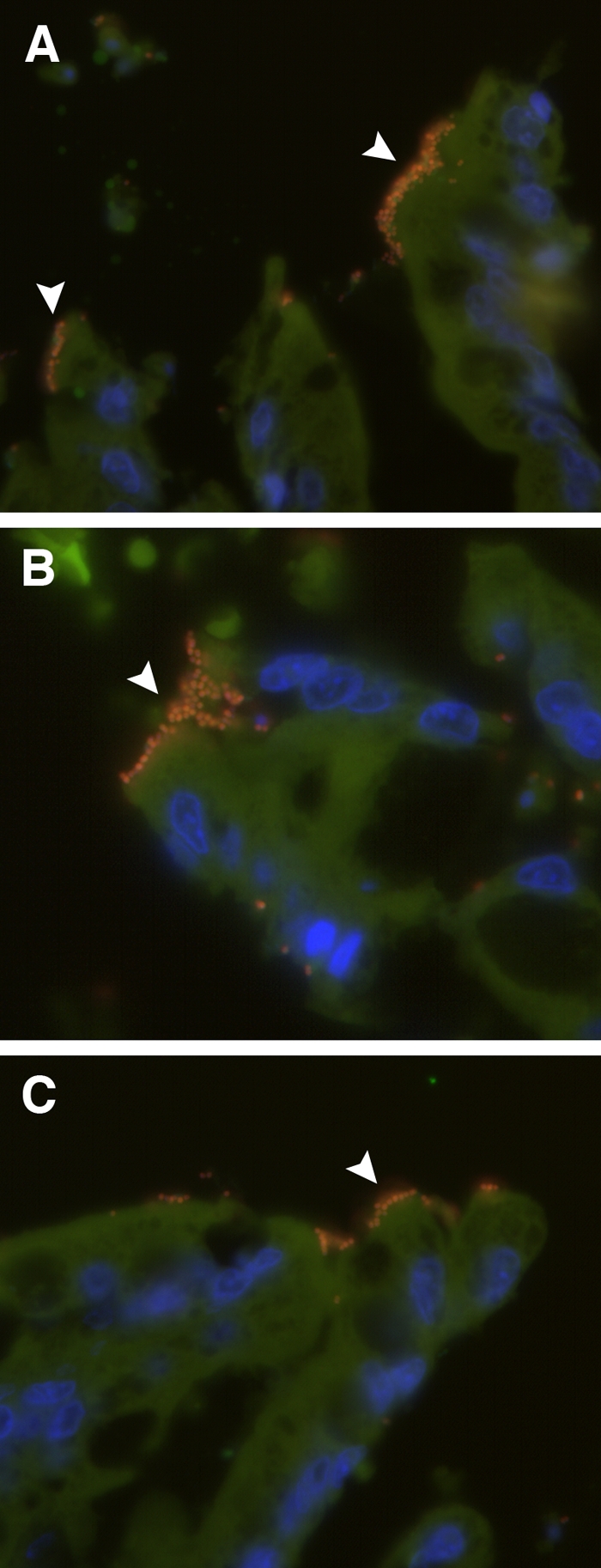
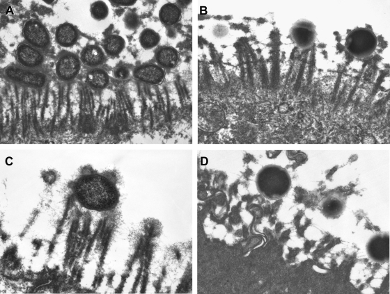
The bacterial causes of diarrhea can be frustrating to identify, and it is likely that many remain undiagnosed. The pathogenic potential of certain bacteria becomes less ambiguous when they are observed to intimately associate with intestinal epithelial cells. In the present study we sought to retrospectively characterize the clinical, in situ molecular, and histopathological features of enteroadherent bacteria in seven unrelated kittens that were presumptively diagnosed with enteropathogenic Escherichia coli (EPEC) on the basis of postmortem light microscopic and, in some cases, microbiological examination. Characterization of the enteroadherent bacteria in each case was performed by Gram staining, in situ hybridization using fluorescence-labeled oligonucleotide probes, PCR amplification of species-specific gene sequences, and ultrastructural imaging applied to formalin-fixed paraffin-embedded sections of intestinal tissue. In only two kittens was EPEC infection confirmed. In the remaining five kittens, enteroadherent bacteria were identified as Enterococcus spp. The enterococci were further identified as Enterococcus hirae on the basis of PCR amplification of DNA extracted from the formalin-fixed, paraffin-embedded tissue and amplified by using species-specific primers. Transmission electron microscopy of representative lesions from E. coli- and Enterococcus spp.-infected kittens revealed coccobacilli adherent to intestinal epithelial cells without effacement of microvilli or cup-and-pedestal formation. Enterococci were not observed, nor were DNA sequences amplified from intestinal tissue obtained from age-matched kittens euthanized for reasons unrelated to intestinal disease. These studies suggest that E. hirae may be a common cause of enteroadherent bacterial infection in pre-weaning-age kittens and should be considered in the differential diagnosis of bacterial disease in this population.
Figures
Similar articles
-
Mortality in kittens is associated with a shift in ileum mucosa-associated enterococci from Enterococcus hirae to biofilm-forming Enterococcus faecalis and adherent Escherichia coli.J Clin Microbiol. 2013 Nov;51(11):3567-78. doi: 10.1128/JCM.00481-13. Epub 2013 Aug 21. J Clin Microbiol. 2013. PMID: 23966487 Free PMC article.
-
Neonatal piglet diarrhoea associated with enteroadherent Enterococcus hirae.J Comp Pathol. 2014 Aug-Oct;151(2-3):137-47. doi: 10.1016/j.jcpa.2014.04.003. Epub 2014 Jun 7. J Comp Pathol. 2014. PMID: 24915885
-
Association of Atypical Enteropathogenic Escherichia coli with Diarrhea and Related Mortality in Kittens.J Clin Microbiol. 2017 Sep;55(9):2719-2735. doi: 10.1128/JCM.00403-17. Epub 2017 Jun 28. J Clin Microbiol. 2017. PMID: 28659315 Free PMC article.
-
Randomized placebo-controlled trial of feline-origin Enterococcus hirae probiotic effects on preventative health and fecal microbiota composition of fostered shelter kittens.Front Vet Sci. 2022 Nov 17;9:923792. doi: 10.3389/fvets.2022.923792. eCollection 2022. Front Vet Sci. 2022. PMID: 36467638 Free PMC article.
-
Fluorescence in situ hybridization for identification of Tritrichomonas foetus in formalin-fixed and paraffin-embedded histological specimens of intestinal trichomoniasis.Vet Parasitol. 2010 Aug 27;172(1-2):139-43. doi: 10.1016/j.vetpar.2010.04.014. Epub 2010 Apr 18. Vet Parasitol. 2010. PMID: 20447769
Cited by
-
Genomic Characterization of Enterococcus hirae From Beef Cattle Feedlots and Associated Environmental Continuum.Front Microbiol. 2022 Jun 27;13:859990. doi: 10.3389/fmicb.2022.859990. eCollection 2022. Front Microbiol. 2022. PMID: 35832805 Free PMC article.
-
Mink (Neovison vison) kits with pre-weaning diarrhea have elevated serum amyloid A levels and intestinal pathomorphological similarities with New Neonatal Porcine Diarrhea Syndrome.Acta Vet Scand. 2018 Aug 15;60(1):48. doi: 10.1186/s13028-018-0403-7. Acta Vet Scand. 2018. PMID: 30111375 Free PMC article.
-
Mortality in kittens is associated with a shift in ileum mucosa-associated enterococci from Enterococcus hirae to biofilm-forming Enterococcus faecalis and adherent Escherichia coli.J Clin Microbiol. 2013 Nov;51(11):3567-78. doi: 10.1128/JCM.00481-13. Epub 2013 Aug 21. J Clin Microbiol. 2013. PMID: 23966487 Free PMC article.
-
A One Health Perspective for Defining and Deciphering Escherichia coli Pathogenic Potential in Multiple Hosts.Comp Med. 2021 Feb 1;71(1):3-45. doi: 10.30802/AALAS-CM-20-000054. Epub 2021 Jan 8. Comp Med. 2021. PMID: 33419487 Free PMC article. Review.
-
Fluorescent In Situ Hybridization for the Detection of Intracellular Bacteria in Companion Animals.Vet Sci. 2024 Jan 22;11(1):52. doi: 10.3390/vetsci11010052. Vet Sci. 2024. PMID: 38275934 Free PMC article. Review.
References
-
- Cave, T. A., H. Thompson, S. W. Reid, D. R. Hodgson, and D. D. Addie. 2002. Kitten mortality in the United Kingdom: a retrospective analysis of 274 histopathological examinations (1986 to 2000). Vet. Rec. 151:497-501. - PubMed
-
- Cheon, D. S., and C. Chae. 1996. Outbreak of diarrhea associated with Enterococcus durans in piglets. J. Vet. Diagn. Invest. 8:123-124. - PubMed
-
- Collins, J. E., M. E. Bergeland, C. J. Lindeman, and J. R. Duimstra. 1988. Enterococcus (Streptococcus) durans adherence in the small intestine of a diarrheic pup. Vet. Pathol. 25:396-398. - PubMed
-
- Devriese, L. A., J. I. Cruz Colque, P. De Herdt, and F. Haesebrouck. 1992. Identification and composition of the tonsillar and anal enterococcal and streptococcal flora of dogs and cats. J. Appl. Bacteriol. 73:421-425. - PubMed
Publication types
MeSH terms
LinkOut - more resources
Full Text Sources
Molecular Biology Databases
Miscellaneous
