. 2010 Aug 6;285(32):24695-706.
doi: 10.1074/jbc.M110.113977. Epub 2010 Jun 2.

Transcription of the transforming growth factor beta activating integrin beta8 subunit is regulated by SP3, AP-1, and the p38 pathway

Affiliations

Transcription of the transforming growth factor beta activating integrin beta8 subunit is regulated by SP3, AP-1, and the p38 pathway

Jennifer A Markovics et al. J Biol Chem. .

Abstract

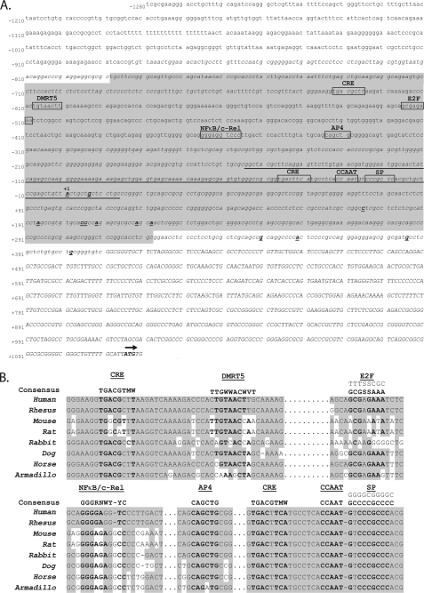
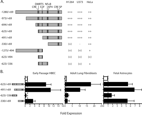
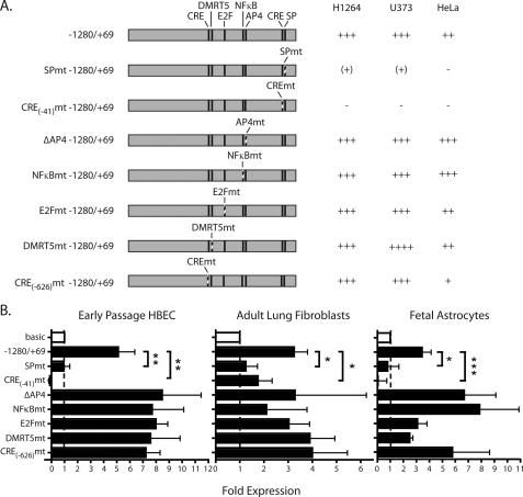
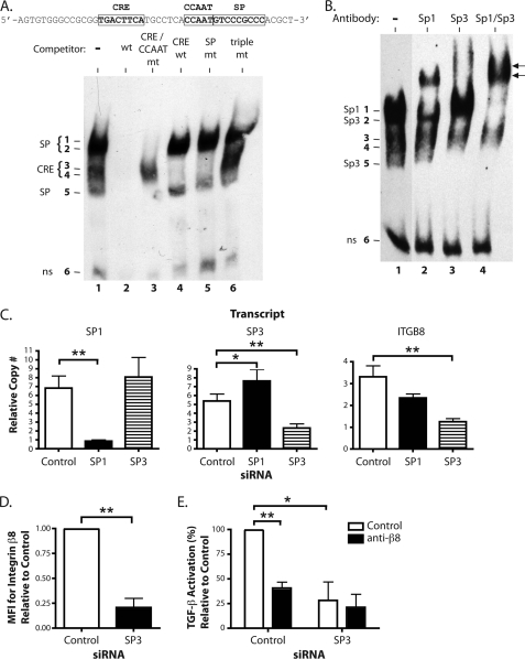
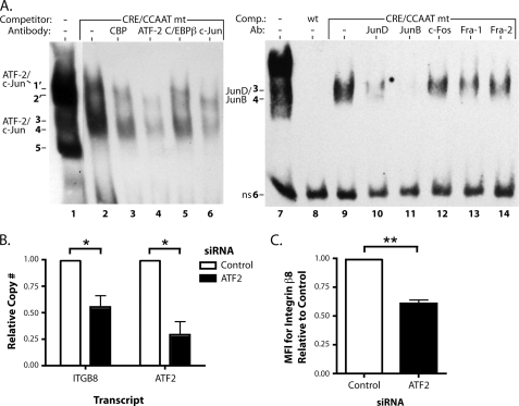
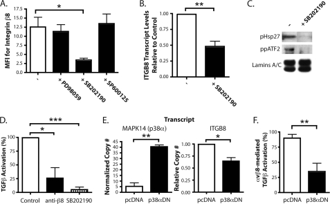
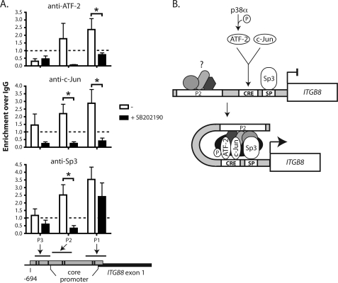
Integrin alphavbeta8 is a critical regulator of transforming growth factor beta activation in vasculogenesis during development, immune regulation, and endothelial/epithelial-mesenchymal homeostasis. Recent studies have suggested roles for integrin beta8 in the pathogenesis of chronic obstructive pulmonary disease, brain arteriovenous malformations, and select cancers (Araya, J., Cambier, S., Markovics, J. A., Wolters, P., Jablons, D., Hill, A., Finkbeiner, W., Jones, K., Broaddus, V. C., Sheppard, D., Barzcak, A., Xiao, Y., Erle, D. J., and Nishimura, S. L. (2007) J. Clin. Invest. 117, 3551-3562; Su, H., Kim, H., Pawlikowska, L., Kitamura, H., Shen, F., Cambier, S., Markovics, J., Lawton, M. T., Sidney, S., Bollen, A. W., Kwok, P. Y., Reichardt, L., Young, W. L., Yang, G. Y., and Nishimura, S. L. (2010) Am. J. Pathol. 176, 1018-1027; Culhane, A. C., and Quackenbush, J. (2009) Cancer Res. 69, 7480-7485; Cambier, S., Mu, D. Z., O'Connell, D., Boylen, K., Travis, W., Liu, W. H., Broaddus, V. C., and Nishimura, S. L. (2000) Cancer Res. 60, 7084-7093). Here we report the first identification and characterization of the promoter for ITGB8. We show that a SP binding site and a cyclic AMP response element (CRE) in the ITGB8 core promoter are required for its expression and that Sp1, Sp3, and several AP-1 transcription factors form a complex that binds to these sites in a p38-dependent manner. Furthermore, we demonstrate the requirement for Sp3, ATF-2, and p38 for the transcription and protein expression of integrin beta8. Additionally, reduction of SP3 or inhibition of p38 blocks alphavbeta8-mediated transforming growth factor beta activation. These results place integrin beta8 expression and activity under the control of ubiquitous transcription factors in a stress-activated and pro-inflammatory pathway.


Figures

FIGURE 1.
FIGURE 1.
Characterization of the 5′ TSS of the ITGB8 gene with surrounding sequence analysis. A, location of the TSS and other sequence features 5′ of the ITGB8 gene. The published ITGB8 5′ untranslated region is indicated in capital letters. The start codon is labeled in bold with an arrow above. The 5′ RACE results from the TSS are labeled in bold and underlined. The most 5′ of these results is labeled as +1 (above), determining the numbering for the presented sequence. Predicted CpG islands are represented in italics. The region of significant homology across 28 mammalian species is underlined. The predicted, overlapping promoter regions are highlighted in light gray. Putative transcription factor binding sites are boxed and labeled above. B, homology of putative transcription factor binding sites of interest in the sequences 5′ of the ITGB8 gene across eight placental mammalian species. Consensus binding sites for the transcription factors are indicated above, the E2F consensus includes both strands in the 5′-3′ orientation. Homologous bases are highlighted in gray.
FIGURE 2.
FIGURE 2.
Identification of the ITGB8 core promoter in cell lines and select primary cells that endogenously express integrin β8. A, ITGB8 reporter constructs and activities in H1264, U373, and HeLa cell lines. −, zero; (+), <2-fold; +, 2–3-fold; ++, >3–8-fold; +++, >8-fold increase in activity, relative to background of the basic reporter construct containing no promoter elements. The constructs are drawn to scale with the location of putative transcription factor binding sites of interest indicated with a line. B, ITGB8 reporter assay results in primary cells, HBEC, adult lung fibroblasts, and fetal astrocytes using select constructs are indicated to the left (± S.E.). Baseline is indicated by a line. The empty bar is baseline, pSEAP basic, whereas filled bars represent relative activity of reporter constructs indicated at the left.
FIGURE 3.
FIGURE 3.
The SP and CRE binding sites are required for ITGB8 promoter activity. A, mutant ITGB8 reporter constructs and activities in H1264, U373, and HeLa cell lines. All constructs are drawn to scale with mutated sites indicated by dashed lines. Fold-induction is as defined as described in the legend to Fig. 2. B, mutant ITGB8 reporter assays in primary HBEC, adult lung fibroblasts, and fetal astrocytes (±S.E.). Baseline is indicated by a line. The empty bar is baseline, pSEAP basic, whereas filled bars represent relative activity of mutant reporter constructs indicated at the left. * = p ≤ 0.05; ** = p ≤ 0.01; *** = p ≤ 0.001.
FIGURE 4.
FIGURE 4.
Sp3 is required for ITGB8 expression and αvβ8-mediated TGF-β activation. A, EMSA in adult lung fibroblasts using a biotinylated probe (sequence indicated above) covering the CRE, CCAAT, and SP transcription factor binding sites in the core promoter with or without the indicated unlabeled competitors. Wt is the unmutated competitor. CRE/CCAAT mt, SP, or triple mt indicates mutations in each or all of the indicated sites. CRE wt is an unrelated CRE consensus site competitor (32). The figure is a composite from a single representative gel. B, EMSA supershift analysis using polyclonal antibodies against Sp1, Sp3, or Sp1 and Sp3 combined. Supershifted complexes are indicated by arrows. The figure is a composite from a single representative gel. C, quantitative RT-PCR results for SP1, SP3, and the ITGB8 in adult lung fibroblasts transfected with siRNA against SP1, SP3, or control (± S.E.). The measured transcript is labeled above each respective graph. D, flow cytometry for surface expression of the integrin β8 subunit on adult lung fibroblasts transfected with siRNA against SP3 or control (± S.E.). MFI, mean fluorescence intensity. E, TGF-β activation assays of adult lung fibroblasts treated with siRNA against SP3 or control using control or anti-β8 blocking antibodies (± S.E.). * = p ≤ 0.05; ** = p ≤ 0.01; *** = p ≤ 0.001.
FIGURE 5.
FIGURE 5.
AP-1 complexes interact with the ITGB8 promoter and ATF-2 regulates ITGB8 expression. A, EMSA supershift analysis using polyclonal antibodies against CRE binding and AP-1 transcription factors. Addition of the CRE/CCAAT double mutant competitor was necessary to displace the Sp1/Sp3 complexes for easier visualization of bands 3 and 4. Bands co-migrating and not displaced by the CRE/CCAT competitor are indicated by 1′ and 2′. Supershifts in the left panel were performed with antibodies against CBP, ATF-2, C/EBPβ, and c-Jun and on the right panel with antibodies against JunD, JunB, c-Fos, Fra-1, and Fra-2. Each panel is a composite from a single representative gel. B, quantitative RT-PCR for ITGB8 and ATF2 from adult lung fibroblasts transfected with an siRNA against ATF2 (± S.E.). C, flow cytometry for surface levels of integrin β8 on adult lung fibroblasts transfected with the same siRNA against ATF2 as in B (± S.E.). MFI, mean fluorescence intensity. * = p ≤ 0.05; ** = p ≤ 0.01.
FIGURE 6.
FIGURE 6.
p38 regulates ITGB8 expression and αvβ8-mediated TGF-β activation. A, flow cytometry for integrin β8 on adult lung fibroblasts treated with MAPK inhibitors, PD98059 (ERK), SB202190 (p38), and SP600125 (JNK) (± S.E.). MFI, mean fluorescence intensity. B, quantitative RT-PCR results for ITGB8 expression in adult lung fibroblasts treated with SB202190, normalized to GAPDH and β-actin and relative to control (± S.E.). C, immunoblot for phosphorylated HSP 27 and dual-phosphorylated ATF-2 from nuclear extracts from adult lung fibroblasts treated ± SB202190. Immunoblot for the nuclear localized proteins, lamins A and C, was used as a loading control. D, TGF-β activation assays of adult lung fibroblasts treated with anti-β8 blocking antibodies or SB202190 (± S.E.). E, quantitative RT-PCR results for MAPK14 (p38α) and ITGB8 in adult lung fibroblasts transfected with plasmids expressing a p38α dominant-negative isoform (p38αDN) or the empty vector control, pcDNA (± S.E.). The measured transcript is labeled above each respective graph. F, TGF-β activation assays of adult lung fibroblasts transfected with plasmids expressing a p38α dominant-negative isoform (p38αDN) or the empty vector control, pcDNA. Percentage (%) of αvβ8-mediated TGF-β activation shown (± S.E.). * = p ≤ 0.05; ** = p ≤ 0.01; *** = p ≤ 0.001.
FIGURE 7.
FIGURE 7.
ATF-2, c-Jun, and Sp3 association with the ITGB8 core promoter requires p38 signaling. A, chromatin immunoprecipitation of ATF-2, c-Jun, or Sp3 in adult lung fibroblasts treated with the p38 inhibitor, SB202190, with PCR amplification of regions from the ITGB8 promoter (± S.E.). Genomic locations of the amplicated regions are indicated in the schematic below the graphs. Essentially identical results were obtained using antibodies against phospho-ATF-2 and total ATF-2 and thus, results were pooled. B, hypothetical model for regulation of the ITGB8 promoter by p38, ATF-2, c-Jun, and Sp3. p38 phosphorylates ATF-2, which heterodimerizes with c-Jun to bind to the CRE site in the ITGB8 core promoter. Along with Sp3, which is already bound to its cognate SP site adjacent to the CRE site in the ITGB8 core promoter, these transcription factors form a higher-order chromatin complex that interacts either directly with the P2 region or indirectly through yet unidentified transcription factors to activate transcription of ITGB8. * = p ≤ 0.05.

Similar articles

Cited by

References

    1. Araya J., Cambier S., Markovics J. A., Wolters P., Jablons D., Hill A., Finkbeiner W., Jones K., Broaddus V. C., Sheppard D., Barzcak A., Xiao Y., Erle D. J., Nishimura S. L. (2007) J. Clin. Invest. 117, 3551–3562 - PMC - PubMed
    1. Su H., Kim H., Pawlikowska L., Kitamura H., Shen F., Cambier S., Markovics J., Lawton M. T., Sidney S., Bollen A. W., Kwok P. Y., Reichardt L., Young W. L., Yang G. Y., Nishimura S. L. (2010) Am. J. Pathol. 176, 1018–1027 - PMC - PubMed
    1. Culhane A. C., Quackenbush J. (2009) Cancer Res. 69, 7480–7485 - PMC - PubMed
    1. Cambier S., Mu D. Z., O'Connell D., Boylen K., Travis W., Liu W. H., Broaddus V. C., Nishimura S. L. (2000) Cancer Res. 60, 7084–7093 - PubMed
    1. Danen E. H., Sonnenberg A. (2003) J. Pathol. 201, 632–641 - PubMed

Publication types

MeSH terms

Substances