. 2010 Aug 6;285(32):24457-65.
doi: 10.1074/jbc.M110.132407. Epub 2010 Jun 2.

DNA polymerase beta ribonucleotide discrimination: insertion, misinsertion, extension, and coding

Affiliations

DNA polymerase beta ribonucleotide discrimination: insertion, misinsertion, extension, and coding

Nisha A Cavanaugh et al. J Biol Chem. .

Abstract

DNA polymerases must select nucleotides that preserve Watson-Crick base pairing rules and choose substrates with the correct (deoxyribose) sugar. Sugar discrimination represents a great challenge because ribonucleotide triphosphates are present at much higher cellular concentrations than their deoxy-counterparts. Although DNA polymerases discriminate against ribonucleotides, many therapeutic nucleotide analogs that target polymerases have sugar modifications, and their efficacy depends on their ability to be incorporated into DNA. Here, we investigate the ability of DNA polymerase beta to utilize nucleotides with modified sugars. DNA polymerase beta readily inserts dideoxynucleoside triphosphates but inserts ribonucleotides nearly 4 orders of magnitude less efficiently than natural deoxynucleotides. The efficiency of ribonucleotide insertion is similar to that reported for other DNA polymerases. The poor polymerase-dependent insertion represents a key step in discriminating against ribonucleotides because, once inserted, a ribonucleotide is easily extended. Likewise, a templating ribonucleotide has little effect on insertion efficiency or fidelity. In contrast to insertion and extension of a ribonucleotide, the chemotherapeutic drug arabinofuranosylcytosine triphosphate is efficiently inserted but poorly extended. These results suggest that the sugar pucker at the primer terminus plays a crucial role in DNA synthesis; a 3'-endo sugar pucker facilitates nucleotide insertion, whereas a 2'-endo conformation inhibits insertion.


Figures

FIGURE 1.
FIGURE 1.
DNA polymerase β nucleoside triphosphate binding pocket and key protein-nucleic acid interactions. A, dNTP binding pocket is composed of nucleic acid (primer terminus and templating base, nt, yellow) and protein (purple). The incoming nucleotide, 2′-deoxyuridine-5′-[(α,β)-imido] triphosphate, is shown hydrogen-bonded (green dashed lines) with the templating nucleotide (dA). The two active site Mg2+ ions are illustrated as light blue spheres. B, stacking of the nascent base pair with the primer terminus positions O3′ of the primer terminus for optimal attack on the α-phosphate of the incoming nucleotide. Two α-helixes (M and N) provide key interactions with the sugar and base moieties of the incoming nucleotide. In addition, Arg-183 (R183) and O3′ of the incoming nucleotide hydrogen bond to a nonbridging oxygen on the β-phosphate (dashed green lines). C, Asp-276 (D276) of α-helix N is positioned above the sugar ring and approaches C2′. The incoming nucleotide is represented as a semi-transparent surface (gray) with C2′ highlighted in pink. The side chain of Asp-276 is also represented as a surface (purple) just above C2′ and would potentially block araCTP that has a hydroxyl at this position. D, primer terminal base pair is highlighted (yellow), and the other bases are gray. The backbone (rather than the side chain) of Tyr-271 (Y271) of α-helix M would potentially block a ribonucleotide with a hydroxyl at C2′ (magenta). Additionally, the side chain of Tyr-271 hydrogen bonds with the minor groove edge of the primer terminal base (dashed green line). Arg-283 (R283) of α-helix N hydrogen bonds with the nucleotide opposite the primer terminus, (n − 1)t, at O4′ of the sugar ring.
FIGURE 2.
FIGURE 2.
Substrates used in this study. A, schematic and sequence of single-nucleotide gapped DNA substrates. DNA substrates were constructed as described under “Experimental Procedures.” The 5′ terminus of the primer strand was radioactively labeled with [γ-32P]ATP. The specific identities of X, Y, and Z are provided in each figure or table. B, structures and abbreviations of modified nucleotide sugars.
FIGURE 3.
FIGURE 3.
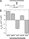
DNA polymerase β sugar discrimination. A, schematic diagram of the single-nucleotide gapped DNA (template dG). B, log plot of catalytic efficiencies for modified sugar substrates. DNA polymerase β misinserts dTTP (wrong base/correct sugar) much less efficiently than ddCTP, rCTP, and araCTP (correct bases/wrong sugars). The base line represents a catalytic efficiency of 10−2 s−1μm−1. The efficiencies are tabulated in Table 1.
FIGURE 4.
FIGURE 4.
Extension of an rNMP-terminated primer by pol β. A, schematic diagram of the single-nucleotide gapped DNA substrates (template dG). The 3′-primer terminus was either dC (left diagram) or rC (right diagram). B, log plot comparing catalytic efficiencies for correct incorporation (dCTP), misinsertion (dTTP), and incorporation of an incorrect sugar (rCTP) using dC- (open bars) or rC-terminated primers (gray-filled bars). The base line represents a catalytic efficiency of 10−2 s−1 μm−1. The efficiencies are tabulated in Tables 1 and 2.
FIGURE 5.
FIGURE 5.
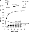
Extension of an araCMP-terminated primer by pol β. A, schematic diagram of the two-nucleotide gapped DNA substrate (see Fig. 2; X has been deleted, Y = dG, and Z = dT). Enzyme (2 nm) was incubated with 200 nm DNA and 35 μm araCTP to fully extend the 5′-32P-labeled primer (>90% in 2.5 min), creating a single-nucleotide gapped substrate with dT in the gap. The rate of extension of the araC-terminated primer was measured in the presence of increasing amounts of dATP (0.5–50 μm). B, dATP concentration dependence of the rate was fit to the Michaelis-Menten equation (solid line; kcat = 3.6 × 10⁻² s−1; Km = 4.1 μm; kcat/Km = 8.8 × 10⁻³ s−1 μm−1). Inset, gel showing 5′-32P-labeled primer (P), araC-terminated primer (P + araC), and final product (P + araC + dA). araCTP was added to all samples; the negative control (C) contained no pol β, and the positive control (N) contained no dATP indicating that nearly all of the primer was extended in the presence of araCTP and that araCTP was not misinserted opposite dT.
FIGURE 6.
FIGURE 6.
Extension of hybrid (rG-dC) or RNA (rG-rC) template-primer termini by pol β. A, schematic diagram of single-nucleotide gapped DNA substrates (templating dG) with a deoxynucleotide (template-primer, dG-dC; left diagram), hybrid (rG-dC; middle diagram), or RNA (rG-rC; right diagram) primer terminus. B, log plot comparing catalytic efficiencies for correct incorporation (dCTP), misinsertion (dTTP), and incorporation of an incorrect sugar (rCTP) using one of the template-primer termini as follows: dG-dC termini (open bars), rG-dC termini (light gray-filled bars), or rG-rC termini (dark gray-filled bars). The base line represents a catalytic efficiency of 10−2 s−1 μm−1. The efficiencies are tabulated in Tables 1 and 2.
FIGURE 7.
FIGURE 7.
Nucleotide incorporation by pol β opposite a templating (coding) ribonucleotide (rG). A, schematic diagram of DNA single-nucleotide gapped DNA substrates with either a templating deoxynucleotide (left diagram, dG) or ribonucleotide (right diagram, rG). B, log plot comparing catalytic efficiencies for correct incorporation (dCTP), misinsertion (dTTP), and incorporation of an incorrect sugar (rCTP) using either the dG (open bars) or the rG template (gray-filled bars). The base line represents a catalytic efficiency of 10−2 s−1 μm−1. The efficiencies are tabulated in Tables 1 and 2.

References

    1. Ferraro P., Franzolin E., Pontarin G., Reichard P., Bianchi V. (2010) Nucleic Acids Res. 38, e85. - PMC - PubMed
    1. Traut T. W. (1994) Mol. Cell. Biochem. 140, 1–22 - PubMed
    1. Astatke M., Grindley N. D., Joyce C. M. (1998) J. Mol. Biol. 278, 147–165 - PubMed
    1. Astatke M., Ng K., Grindley N. D., Joyce C. M. (1998) Proc. Natl. Acad. Sci. U.S.A. 95, 3402–3407 - PMC - PubMed
    1. Gardner A. F., Joyce C. M., Jack W. E. (2004) J. Biol. Chem. 279, 11834–11842 - PubMed

Publication types

LinkOut - more resources