. 2010 Jul 30;285(31):23916-24.
doi: 10.1074/jbc.M110.127936. Epub 2010 Jun 2.

Serine residues in the cytosolic tail of the T-cell antigen receptor alpha-chain mediate ubiquitination and endoplasmic reticulum-associated degradation of the unassembled protein

Affiliations

Serine residues in the cytosolic tail of the T-cell antigen receptor alpha-chain mediate ubiquitination and endoplasmic reticulum-associated degradation of the unassembled protein

Shuhei Ishikura et al. J Biol Chem. .

Abstract

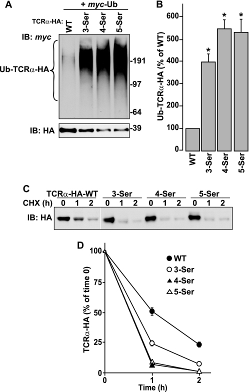
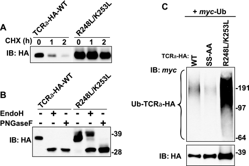
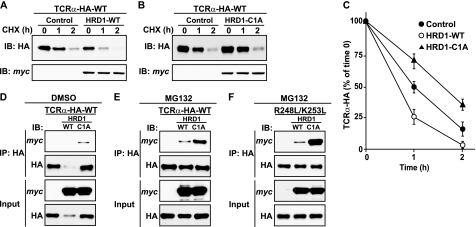
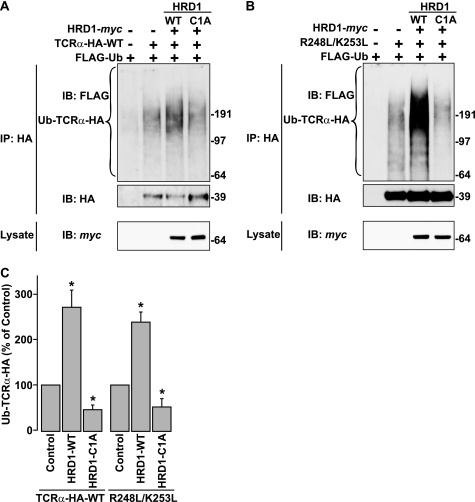
The T-cell antigen receptor (TCR) alpha-chain (TCRalpha) is a type I integral membrane protein that becomes ubiquitinated and targeted to the endoplasmic reticulum (ER)-associated degradation (ERAD) pathway when it fails to assemble into the heteromeric TCR complex. Remarkably, TCRalpha has a cytosolic tail of only five amino acid residues (i.e. RLWSS), none of which is the conventional ubiquitin acceptor, lysine. Herein we report that substitution of two conserved serine residues in the cytosolic tail of TCRalpha to alanine decreased ubiquitination, whereas placement of additional serine residues enhanced it. Moreover, replacement of the cytosolic serine residues by other ubiquitinatable residues (i.e. cysteine, threonine, or lysine) allowed ubiquitination to take place. Serine-dependent ubiquitination perfectly correlated with targeting of TCRalpha for ERAD. We also found that this ubiquitination was mediated by the ER-localized ubiquitin ligase, HRD1. These findings indicate that serine-dependent, HRD1-mediated ubiquitination targets TCRalpha to the ERAD pathway.


Figures

FIGURE 1.
FIGURE 1.
Mutation of two serine residues in the cytosolic tail of TCRα inhibits its ubiquitination and degradation. A, amino acid sequence of the transmembrane domain and cytosolic tail of WT TCRα and its mutants. A shortened HA tag (YPYDVPDYA) was appended to the C terminus of all constructs. Substituted residues are shown in red. B, HeLa cells transfected with plasmids encoding the indicated TCRα-HA constructs with or without Myc-Ub were left untreated or treated with 50 μm MG132 for 4 h at 37 °C. After lysis in RIPA buffer containing a protease inhibitors mixture, N-ethylmaleimide and iodoacetamide, TCRα-HA was immunoprecipitated with anti-HA monoclonal antibody. Immunoprecipitates were analyzed by SDS-PAGE and immunoblotting (IB) with anti-HA and anti-Myc polyclonal antibodies. Molecular masses of marker proteins (in kilodaltons) are indicated on the right. C, levels of ubiquitinated TCRα-HA were quantified by densitometry, normalized to levels of non-ubiquitinated TCRα-HA, and expressed as percent of WT TCRα-HA levels. Values represent the mean ± S.E. from three independent experiments. *, p < 0.05 versus WT. D, HeLa cells were transfected with plasmids encoding the indicated TCRα-HA constructs. After 24 h, the cells were left untreated or treated with 100 μg/ml CHX for the indicated time periods. Cell lysates were prepared in RIPA buffer, and analyzed by SDS-PAGE and immunoblotting with an anti-HA monoclonal antibody. E, TCRα-HA remaining at each time point was quantified and expressed as percent of TCRα present at time 0. Values represent the mean ± S.E. from three independent experiments.
FIGURE 2.
FIGURE 2.
Addition of serine residues to the cytosolic tail of TCRα enhances its ubiquitination and degradation. A and B, ubiquitination of WT TCRα and serine-addition mutants of TCRα (Fig. 1A) was determined as described in the legend to Fig. 1, B and C. *, p < 0.05 versus WT. C and D, CHX chase experiments and quantification were performed as described in the legend to Fig. 1, D and E.
FIGURE 3.
FIGURE 3.
Ubiquitination and degradation of TCRα mutants bearing single serine residues at different positions within the cytosolic tail. A and B, ubiquitination of WT TCRα and single-serine insertion mutants of TCRα (Fig. 1A) was determined as described in the legend to Fig. 1, B and C. *, p < 0.05 versus SS-AA mutant. C and D, CHX chase experiments and quantification were performed as described in the legend to Fig. 1, D and E.
FIGURE 4.
FIGURE 4.
Ubiquitination and degradation of TCRα bearing cysteine, threonine, or lysine residues in place of serine residues in the cytosolic tail. A and B, ubiquitination of WT TCRα and cysteine, threonine, or lysine substitution mutants of TCRα (Fig. 1A) was determined as described in the legend to Fig. 1, B and C. *, p < 0.05 versus WT. C and D, CHX chase experiments and quantification were performed as described in the legend to Fig. 1, D and E.
FIGURE 5.
FIGURE 5.
Ubiquitination is independent of two basic residues in the transmembrane domain of TCRα. A, CHX chase experiments were performed as described in the legend to Fig. 1D. B, lysates from HeLa cells transfected with plasmids encoding WT TCRα-HA or R248L/K253L mutant were left untreated or digested with Endo H or PNGase F before immunoblotting with anti-HA monoclonal antibody. C, ubiquitination of WT and mutant TCRα constructs (Fig. 1A) was determined as described in the legend to Fig. 1B. Molecular mass markers (in kilodaltons) are indicated on the right.
FIGURE 6.
FIGURE 6.
HRD1 Ub ligase interacts with TCRα and promotes its degradation. A–C, HeLa cells were transfected with plasmids encoding WT TCRα-HA with or without WT or C1A mutant HRD1. CHX chase experiments and quantification were performed as described in the legend to Fig. 1, D and E. D–F, HeLa cells transfected with plasmids encoding WT TCRα-HA or R248L/K253L mutant with or without WT or C1A mutant HRD1 were left untreated or treated with 50 μm MG132 for 4 h at 37 °C. After lysis, TCRα-HA was immunoprecipitated with anti-HA monoclonal antibody. Lysates (5% input) and immunoprecipitates (IP) were analyzed by SDS-PAGE and immunoblotting (IB) with anti-HA and anti-Myc polyclonal antibodies.
FIGURE 7.
FIGURE 7.
HRD1 Ub ligase mediates TCRα ubiquitination. A and B, HeLa cells were transfected with the indicated constructs. Ubiquitination of TCRα-HA was determined in the absence of MG132 as described in the legend to Fig. 1B with the exception of using anti-FLAG antibody instead of anti-Myc polyclonal antibody to detect ubiquitinated TCRα-HA. C, ubiquitinated TCRα-HA levels were normalized to non-ubiquitinated TCRα-HA levels. Values represent the mean ± S.E. from three independent experiments. *, p < 0.05 versus untreated with HRD1 cDNA (Control).
FIGURE 8.
FIGURE 8.
HRD1 knockdown inhibits degradation and ubiquitination of TCRα. A and B, HeLa cells were left untreated (UnTransfect) or treated with non-targeting siRNA (siControl) or HRD1 siRNA (siHRD1). After 72 h, cells were transfected with WT TCRα-HA and then CHX chase experiments and quantification were performed as described in the legend to Fig. 1, D and E. Expression of endogeneous HRD1 was determined by immunoblotting with anti-HRD1 polyclonal antibody. C, HeLa cells treated with the indicated siRNA were transfected with FLAG-Ub, and WT TCRα-HA or R248L/K253L mutant. Ubiquitination of TCRα-HA was determined in the absence of MG132 as described in the legend to Fig. 7. Endogeneous HRD1 in the lysate was detected as described above. D, ubiquitinated TCRα-HA levels were normalized to non-ubiquitinated TCRα-HA levels. Values represent the mean ± S.E. from three independent experiments. *, p < 0.05 versus treated with non-targeting siRNA (siControl).

Similar articles

Cited by

References

    1. Ellgaard L., Helenius A. (2003) Nat. Rev. Mol. Cell Biol. 4, 181–191 - PubMed
    1. Kleizen B., Braakman I. (2004) Curr. Opin. Cell Biol. 16, 343–349 - PubMed
    1. Kostova Z., Wolf D. H. (2003) EMBO J. 22, 2309–2317 - PMC - PubMed
    1. Meusser B., Hirsch C., Jarosch E., Sommer T. (2005) Nat. Cell Biol. 7, 766–772 - PubMed
    1. Vembar S. S., Brodsky J. L. (2008) Nat. Rev. Mol. Cell Biol. 9, 944–957 - PMC - PubMed

Publication types

MeSH terms

LinkOut - more resources