. 2010 Aug 6;285(32):24665-75.
doi: 10.1074/jbc.M109.091256. Epub 2010 Jun 2.

Plasminogen activator inhibitor-1 is a transcriptional target of the canonical pathway of Wnt/beta-catenin signaling

Affiliations

Plasminogen activator inhibitor-1 is a transcriptional target of the canonical pathway of Wnt/beta-catenin signaling

Weichun He et al. J Biol Chem. .

Abstract

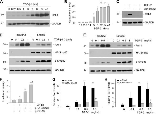
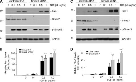
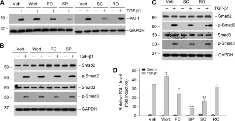
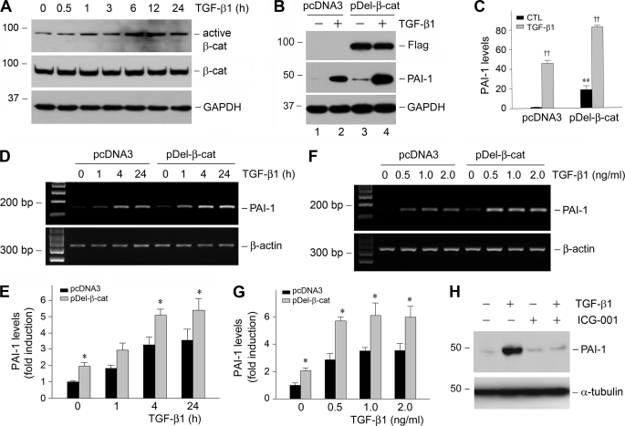
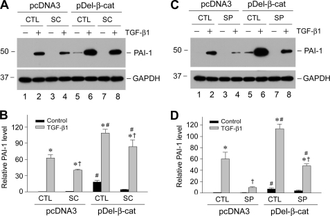
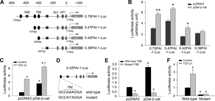
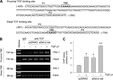
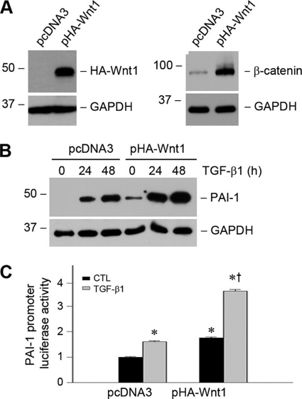
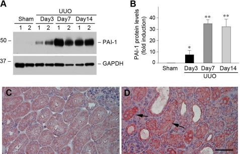
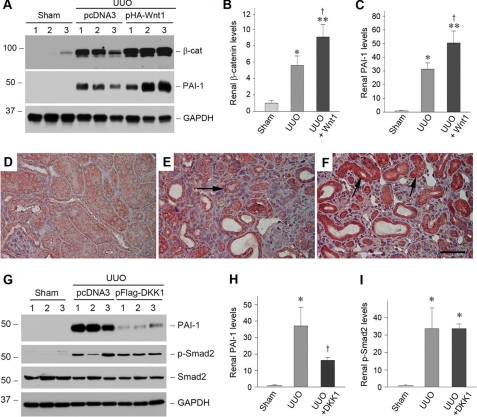
Plasminogen activator inhibitor-1 (PAI-1) is a multifunctional glycoprotein that plays a critical role in the pathogenesis of chronic kidney and cardiovascular diseases. Although transforming growth factor (TGF)-beta1 is a known inducer of PAI-1, how it controls PAI-1 expression remains enigmatic. Here we investigated the mechanism underlying TGF-beta1 regulation of PAI-1 in kidney tubular epithelial cells (HKC-8). Surprisingly, overexpression of Smad2 or Smad3 in HKC-8 cells blocked PAI-1 induction by TGF-beta1, whereas knockdown of them sensitized the cells to TGF-beta1 stimulation, suggesting that Smad signaling is not responsible for PAI-1 induction. Blockade of several TGF-beta1 downstream pathways such as p38 MAPK or JNK, but not phosphatidylinositol 3-kinase/Akt and ERK1/2, only partially inhibited PAI-1 expression. TGF-beta1 stimulated beta-catenin activation in tubular epithelial cells, and ectopic expression of beta-catenin induced PAI-1 expression, whereas inhibition of beta-catenin abolished its induction. A functional T cell factor/lymphoid enhancer-binding factor-binding site was identified in the promoter region of the PAI-1 gene, which interacted with T cell factor upon beta-catenin activation. Deletion or site-directed mutation of this site abolished PAI-1 response to beta-catenin or TGF-beta1 stimulation. Similarly, ectopic expression of Wnt1 also activated PAI-1 expression and promoter activity. In vivo, PAI-1 was induced in kidney tubular epithelia in obstructive nephropathy. Delivery of Wnt1 gene activated beta-catenin and promoted PAI-1 expression after obstructive injury, whereas blockade of Wnt/beta-catenin signaling by Dickkopf-1 gene inhibited PAI-1 induction. Collectively, these studies identify PAI-1 as a direct downstream target of Wnt/beta-catenin signaling and demonstrate that PAI-1 induction could play a role in mediating the fibrogenic action of this signaling.


Figures

FIGURE 1.
FIGURE 1.
Smad blocks PAI-1 expression induced by TGF-β1 in proximal tubular epithelial cells. A and B, TGF-β1 induced PAI-1 expression in a time-dependent manner. Human proximal tubular epithelial cells (HKC-8) were treated with TGF-β1 (2 ng/ml) for various periods of time as indicated. Shown in A is the representative Western blot result. Quantitative determination of PAI-1 induction after normalization with GAPDH is presented in B. C, TGF-β receptor signaling is required for PAI-1 induction. HKC-8 cells were pretreated with activin receptor-like kinase-5 inhibitor SB431542 (10 μm), followed by incubation with TGF-β1 (2 ng/ml) for 24 h as indicated. D and E, overexpression of Smad2 or Smad3 inhibited PAI-1 induction in response to TGF-β1 stimulation. HKC-8 cells were transiently transfected with HA-tagged Smad2 or Smad3, or empty pcDNA3 vector, followed by incubation with different doses of TGF-β1 as indicated. Whole cell lysates were immunoblotted with specific antibodies against PAI-1, HA, phospho-Smad2, phospho-Smad3, and GAPDH, respectively. F, Smad3 promotes the SBE-driven luciferase reporter activity. HKC-8 cells were cotransfected with SBE4-Luc reporter plasmid and Smad3 expression vector (pHA-Smad3) or empty vector (pcDNA3), followed by incubation with TGF-β1 for 48 h as indicated. Relative luciferase activities (arbitrary unit) were reported. G and H, graphic presentations of PAI-1 abundance after transfection with Smad2 or Smad3 are shown. The data are presented as the means ± S.E. of three experiments. *, p < 0.05; **, p < 0.01 versus controls. †, p < 0.05; ††, p < 0.01 versus pcDNA3.
FIGURE 2.
FIGURE 2.
Knockdown of Smad2 or Smad3 enhances PAI-1 induction after TGF-β1 stimulation. HKC-8 cells were transiently transfected with control (Cont.) siRNA, Smad2, or Smad3 siRNA (120 nm) and then treated with different doses of TGF-β1 as indicated. Whole cell lysates were immunoblotted with specific antibodies against PAI-1, Smad2, Smad3, phospho-Smad2, phospho-Smad3, and GAPDH, respectively. Representative Western blots (A and C) and the relative abundance of PAI-1 after normalization with GAPDH (B and D) are presented. The data (relative to the controls = 1.0) are presented as the means ± S.E. of three experiments. *, p < 0.05; **, p < 0.01 versus controls. †, p < 0.01 versus control siRNA group with TGF-β1.
FIGURE 3.
FIGURE 3.
Activation of MAPK signaling is partially responsible for PAI-1 induction by TGF-β1. HKC-8 cells were pretreated with either various chemical inhibitors or vehicle (Veh., 0.1% Me2SO) as indicated for 30 min, followed by incubation in the absence or presence of TGF-β1 (2 ng/ml) for 24 h. PD, PD98059 (MEK1 inhibitor) (10 μm); wort., wortmannin (PI3K inhibitor) (10 nm); SC, SC68376 (p38 MAPK inhibitor) (20 μm); SP, SP600125 (JNK inhibitor) (20 μm); RO, Ro31-8220 (pan-specific protein kinase C inhibitor) (10 μm). A, representative Western blots of PAI-1 expression after various treatments. B and C, Smad2/3 abundance and phosphorylation in the presence of various inhibitors as indicated. D, quantitative determination of the relative PAI-1 abundance after normalization with GAPDH. The data (relative to the controls = 1.0) are presented as the means ± S.E. of three experiments. *, p < 0.05; **, p < 0.01 versus vehicle controls.
FIGURE 4.
FIGURE 4.
Activation of β-catenin signaling by TGF-β1 mediates PAI-1 induction. A, TGF-β1 induces β-catenin activation in tubular epithelial cells. HKC-8 cells were treated with TGF-β1 (2 ng/ml) for various periods of time as indicated, and the cell lysates were immunoblotted with antibodies against active β-catenin, total β-catenin, and GAPDH, respectively. B and C, ectopic expression of stabilized β-catenin promotes PAI-1 induction at basal and TGF-β1-stimulated conditions. HKC-8 cells stably transfected with either empty vector (pcDNA3) or FLAG-tagged, N-terminal truncated β-catenin (pDel-β-cat) were incubated in the absence or presence of TGF-β1 (2 ng/ml) for 48 h. A representative Western blot (B) and the quantitative data (C) are presented. CTL, control. **, p < 0.01 versus pcDNA3 controls. ††, p < 0.01 versus without TGF-β1 (n = 3). D–G, expression of stabilized β-catenin also promotes PAI-1 mRNA expression. HKC-8 cells stably transfected with either empty vector (pcDNA3) or FLAG-tagged, N-terminal truncated β-catenin (pDel-β-cat) were incubated in the absence or presence of TGF-β1 for various periods of time (D and E) at different concentrations (F and G). The quantitative data on PAI-1 mRNA induction (fold over the controls) are presented in E and G. *, p < 0.05 versus pcDNA3 controls. H, inhibition of β-catenin signaling blocked TGF-β1-induced PAI-1 expression. HKC-8 cells were pretreated with a specific small molecule β-catenin inhibitor (ICG-001) (10 μm) for 30 min, followed by incubation in the absence or presence of TGF-β1 (2 ng/ml) for 24 h.
FIGURE 5.
FIGURE 5.
Multiple signal pathways downstream of TGF-β1 are involved in mediating PAI-1 induction. HKC-8 cells stably transfected with either empty vector (pcDNA3) or FLAG-tagged, N-terminal truncated β-catenin (pDel-β-cat) were incubated with TGF-β1 (2 ng/ml) in the absence or presence of SC68376 (SC, p38 MAPK inhibitor) (A and B) or SP600125 (SP, JNK inhibitor) (C and D). Representative Western blot (A and C) and quantitative data (B and D) on PAI-1 induction (fold over the controls) are presented. *, p < 0.05 versus controls without TGF-β1. †, p < 0.05 versus controls without inhibitors. #, p < 0.05 versus pcDNA3 controls (n = 3). CTL, control.
FIGURE 6.
FIGURE 6.
PAI-1 gene promoter harbors a functional TBE that is responsible for β-catenin-mediated PAI-1 induction. A, diagram shows the construction of various luciferase reporter plasmids containing different lengths of the promoter region of human PAI-1 gene linked to the coding sequence of luciferase gene. Putative TBE, SBE, Sp1, and AP1 sites are indicated. B, β-catenin activates PAI-1 promoter in tubular epithelial cells. HKC-8 cells were cotransfected with various PAI-1 promoter-luciferase reporter plasmids as shown in A and stabilized β-catenin expression vector (pDel-β-cat) or empty vector (pcDNA3). R. reniformis luciferase vector (pRL-TK) was used as an internal control for transfection efficiency. Luciferase activities (arbitrary unit) were calculated, reported after normalizing for transfection efficiency, and presented as the means ± S.E. of three experiments. *, p < 0.05; **, p < 0.01 versus pcDNA3 controls. C, PAI-1 promoter harbors a functional TBE (0.42PAI-1-Luc) that is responsive to β-catenin and TGF-β1 stimulation. HKC-8 cells were cotransfected with 0.42PAI-1-Luc reporter plasmid with pcDNA3 or pDel-β-cat, followed by incubation without or with TGF-β1 (2 ng/ml) for 48 h. D–F, site-directed mutation in the TBE of PAI-1 promoter abolishes its responsiveness to β-catenin and TGF-β1 stimulation. The construction of wild-type and mutant TBE reporter plasmids (D) and luciferase activities (E and F) are presented. *, p < 0.05 versus controls; †, p < 0.05 versus pDel-β-cat alone.
FIGURE 7.
FIGURE 7.
Activation of β-catenin promotes TCF binding to the proximal TBE in PAI-1 promoter. A, partial sequence of PAI-1 gene promoter region. Bold letters indicate the putative TBE. P1 and P2 indicate the primer pair encompassing the proximal TBE, whereas P3 and P4 indicate the prime pair for the distal TBE. B and C, ChIP assay revealed that activation of β-catenin (either by TGF-β1 or by overexpressing stabilized β-catenin) promoted TCF binding to the proximal TBE but not distal TBE. Representative ChIP assay (B) and quantitative ChIP data (C) are presented. *, p < 0.05 versus controls; **, p < 0.01 versus controls; †, p < 0.05 versus TGF-β1 or pDel-β-cat alone (n = 3).
FIGURE 8.
FIGURE 8.
Ectopic expression of Wnt1, an upstream activator of β-catenin signaling, promotes PAI-1 expression. A, ectopic expression Wnt1 increases β-catenin protein abundance in tubular epithelial cells. HKC-8 cells were stably transfected with HA-tagged Wnt1, and cell lysates were immunoblotted with antibodies against HA and β-catenin, respectively. B, ectopic expression of Wnt1 promotes PAI-1 induction at basal and TGF-β1-stimulated conditions. HKC-8 cells stably transfected with either pcDNA3 or pHA-Wnt1 were incubated in the absence or presence of TGF-β1 (2 ng/ml) for various periods of time as indicated. C, Wnt1 stimulates PAI-1 promoter activities in tubular epithelial cells. HKC-8 cells were cotransfected with PAI-1 promoter-luciferase reporter plasmid (0.42PAI-1-Luc) and Wnt1 expression vector (pHA-Wnt1) or empty vector (pcDNA3), followed by incubation without or with TGF-β1 as indicated. The luciferase activities (arbitrary unit) were calculated, reported after normalizing for transfection efficiency, and presented as the means ± S.E. of three experiments. *, p < 0.05 versus pcDNA3 controls. †, p < 0.05 versus without TGF-β1 (n = 3). CTL, control.
FIGURE 9.
FIGURE 9.
PAI-1 is induced specifically in renal tubular epithelial cells after obstructive injury. A and B, Western blot analyses show PAI-1 induction in the obstructed kidney at different time points after UUO. Representative Western blot (A) and quantitative data after normalization with GAPDH (B) are presented. The data are presented as the means ± S.E. of five animals (n = 5). *, p < 0.05; **, p < 0.01 versus sham controls. C and D, immunohistochemical staining shows the localization of PAI-1 protein in obstructive nephropathy. Kidney sections from sham (C) and UUO (D) were immunostained with antibody against PAI-1. The arrows indicate PAI-1-positive renal tubular epithelial cells. Scale bar, 50 μm.
FIGURE 10.
FIGURE 10.
Wnt/β-catenin signaling modulates PAI-1 expression in vivo. A–C, expression of exogenous Wnt1 gene induces renal β-catenin and PAI-1 expression after obstructive injury. Representative Western blots (A) and relative levels (fold induction over sham controls) of β-catenin (B) and PAI-1 (C) are presented. Numbers 1–3 indicate individual animals in a given group. The data are presented as the means ± S.E. of five animals (n = 5). *, p < 0.05; **, p < 0.01 versus sham controls. †, p < 0.05 versus UUO controls. D–F, immunohistochemical staining shows the localization of PAI-1 protein in different groups. Kidney sections from sham (D), UUO (E), and UUO plus Wnt1 (F) were immunostained with antibody against PAI-1. The arrows indicate positive staining. Scale bar, 50 μm. G–I, blockade of Wnt/β-catenin canonical pathway by DKK1 inhibits renal PAI-1 expression but not Smad2 phosphorylation after obstructive injury. Representative Western blots (G) and relative levels (fold induction over sham controls) of PAI-1 (H) and phospho-Smad2 (I) are presented. The data are presented as the means ± S.E. of three to five animals (n = 3–5). *, p < 0.05 versus sham controls. †, p < 0.05 versus UUO controls.

Similar articles

Cited by

References

    1. Eddy A. A., Fogo A. B. (2006) J. Am. Soc. Nephrol. 17, 2999–3012 - PubMed
    1. Ma L. J., Fogo A. B. (2009) Front. Biosci. 14, 2028–2041 - PMC - PubMed
    1. Nagamine Y. (2008) Thromb. Haemost. 100, 1007–1013 - PubMed
    1. Hamano K., Iwano M., Akai Y., Sato H., Kubo A., Nishitani Y., Uyama H., Yoshida Y., Miyazaki M., Shiiki H., Kohno S., Dohi K. (2002) Am. J. Kidney Dis. 39, 695–705 - PubMed
    1. Paueksakon P., Revelo M. P., Ma L. J., Marcantoni C., Fogo A. B. (2002) Kidney Int. 61, 2142–2148 - PubMed

Publication types